A DOUBLE-PILLAR SUPPORTED ACHROMATIC COMPOUND MICROSCOPE
ENGLISH
c. 1850-1860
Collection of, and Text Written by Dr Jurriaan de Groot
Please Click On Any Picture for a Larger Version
DESCRIPTION:
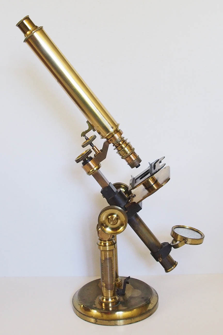
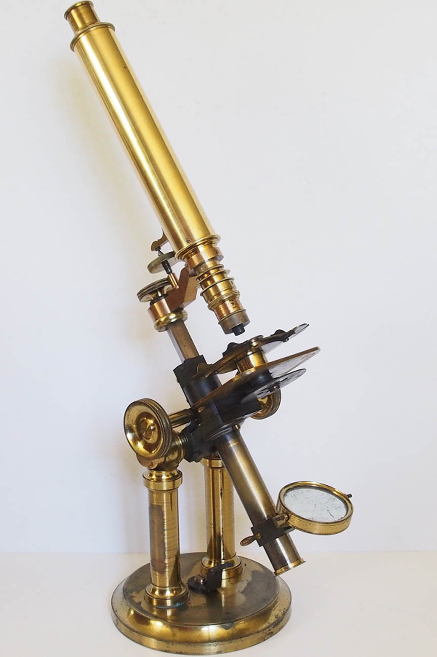
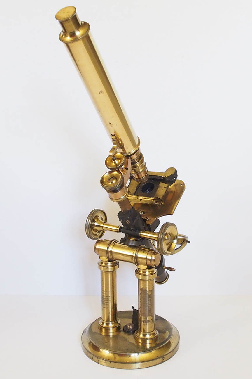
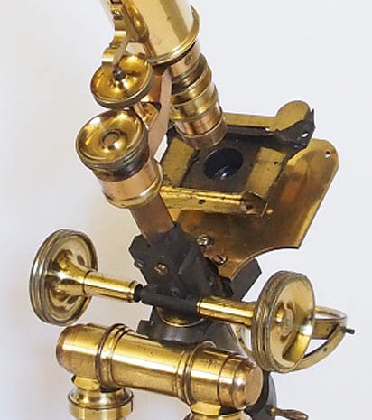
This microscope is supported by a massive brass circular foot 160 mm in diameter, and 22 mm thick. From this arise two large brass pillars 25 mm in diameter and 144 mm high. These carry a horizontal circular support for the body on trunnions, which act as the pivot point for allowing the body to be inclined at angles ranging from vertical to 90 degrees(horizontal). There is an oxidized brass curved stop affixed to the circular base to register the upright position. The short limb is of oxidized brass and arises from the sturdy stage support. Its upper end contains a triangular bar with rack and pinion coarse adjustment which is operated by two knurled brass knobs. The horizontal arm is curved upwards at the body tube end, and attaches to the triangular bar with a knurled round brass nut. The arm also supports the fine adjustment which is activated by a knurled brass head placed at the rear of the body tube and moves a short lever acting on a sprung nose piece. The body tube is 210 mm long and takes eye pieces of a large 36.4 mm diameter. The plain brass stage measures 94 x 97 mm and this supports an accessory stage with a one fixed and one sliding bar to hold slides or accessories. This can be rotated by hand through 360 degrees, and is fitted by means of a central brass sleeve. There is an under stage disc of aperture stops affixed to a short cylinder which screws into the lower part of the stage. The tail piece is an extension of the body and supports a gimballed plano-concave mirror which can be slid up and down the tail piece with two brass lugs. When in use this microscope stands 500 mm tall.
ACCESSORIES:
EYE PIECES:
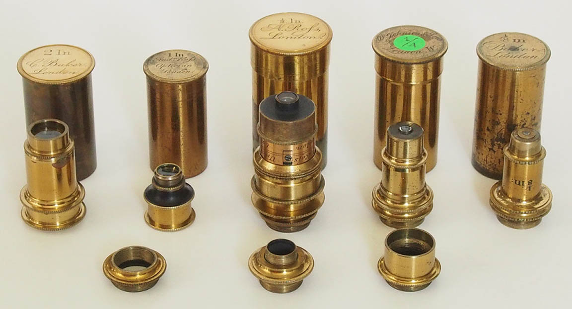
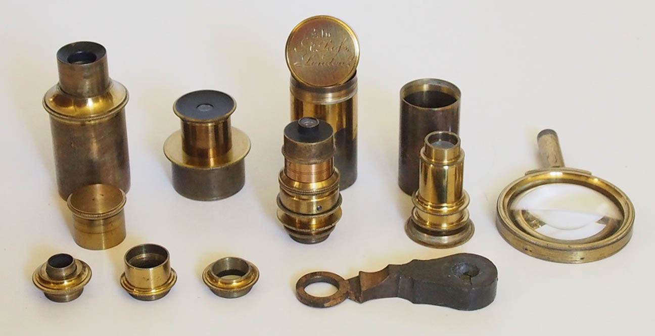
These include an eye piece with dust cap, having an approximate magnification of 5X, and a shorter top hat eye piece of about 10X magnification. Both have an external diameter of 36.4 mm.
OBJECTIVES:
Optical data for these obtained with stage micrometer and Cheshire apertometer:
| Objective |
n.a. | Magnification
with 5 X eyepiece | Magnification
with 10 X eyepiece |
|---|
| 2 In | 0.11 | 35 | 67 |
| 1 In | 0.15 | 60 | 110 |
| 1/2 In | 0.50 | 124 | 225 |
| 1/4 In | 0.60 | 250 | 460 |
| 1/8 In | 0.84 | 500 | 900 |
When acquired, this microscope came with a 2 inch Baker objective with female Ross thread, and a very large 1/2 inch Ross objective with RMS thread. Also present was a set of adapters from RMS to pre-RMS Smith& Beck, and from this to male Ross thread, which -when used together- allow for the use of Ross female internal thread objectives. An early 1 inch Ross objective of this type was added to the set, alongside an unsigned period 1/4 inch, and a Baker 1/8 inch objective, both with RMS thread. Amongst the original accessories was also an adapter for the use of pre-RMS Dancer objectives.
OTHER ACCESSORIES:
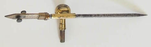
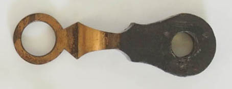 These include a stage forceps(left) and a separate arm for use as a simple dissection microscope(right). This appears to have been bronzed to a crude center support, raising the possibility that this microscope may have originally had a double arm, as in other examples of this model, and was later modified, along with
the fine adjustment arrangement.
These include a stage forceps(left) and a separate arm for use as a simple dissection microscope(right). This appears to have been bronzed to a crude center support, raising the possibility that this microscope may have originally had a double arm, as in other examples of this model, and was later modified, along with
the fine adjustment arrangement.
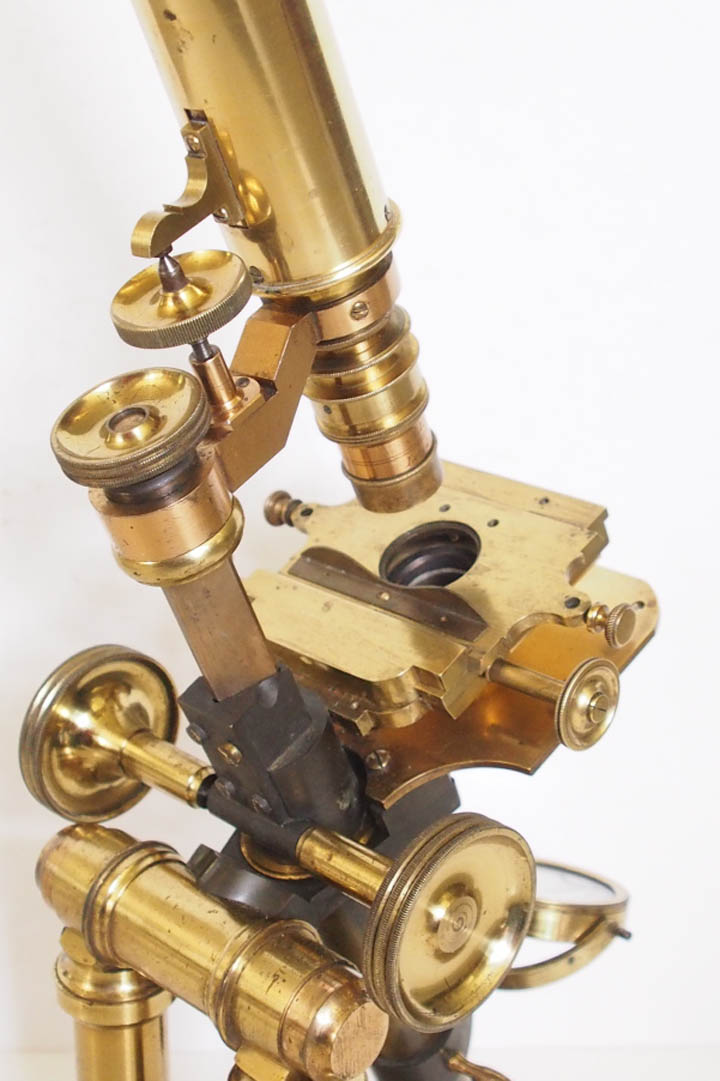 A period mechanical stage was sourced and adapted to be used instead of the original stage, and slots into the cylindrical sleeve of the lower plain stage in a similar fashion. This features horizontally opposed controls for the X and Y axes of movement, and two separate holes into which stage accessories can be fixed with screws.
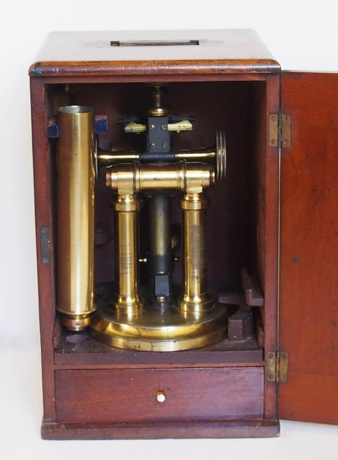
CASE:
The original mahogany case measures 370 mm (H) x 230 mm (W) x 240 mm (D). There is an accessory drawer below the main compartment where the microscope is stored on a sliding square of mahogany, which has a separate recess for the arm and body tube.
CONDITION:
The present owner has re-lacquered the body tube and lower plain stage, given their total loss of lacqer. The rest of the lacquer was left in original condition.
HISTORY AND POSSIBLE ORIGIN OF THIS TYPE OF MICROSCOPE:
Microscopes of double pillar design were made by continental opticians, especially Hartnack and Nachet, while in England James Smith began using this design for his "Large Best" models around 1846. This design was also popular in the USA, with Zentmayer, Bulloch, Bausch & Lomb,and Tolles microscopes using double
pillar supports for their larger models. Thomas Ross followed suit in 1879 with his Ross-Zentmayer model. However, most of these designs featured a horseshoe, or flat tripod base, and not the type of heavy circular foot as in this instrument.
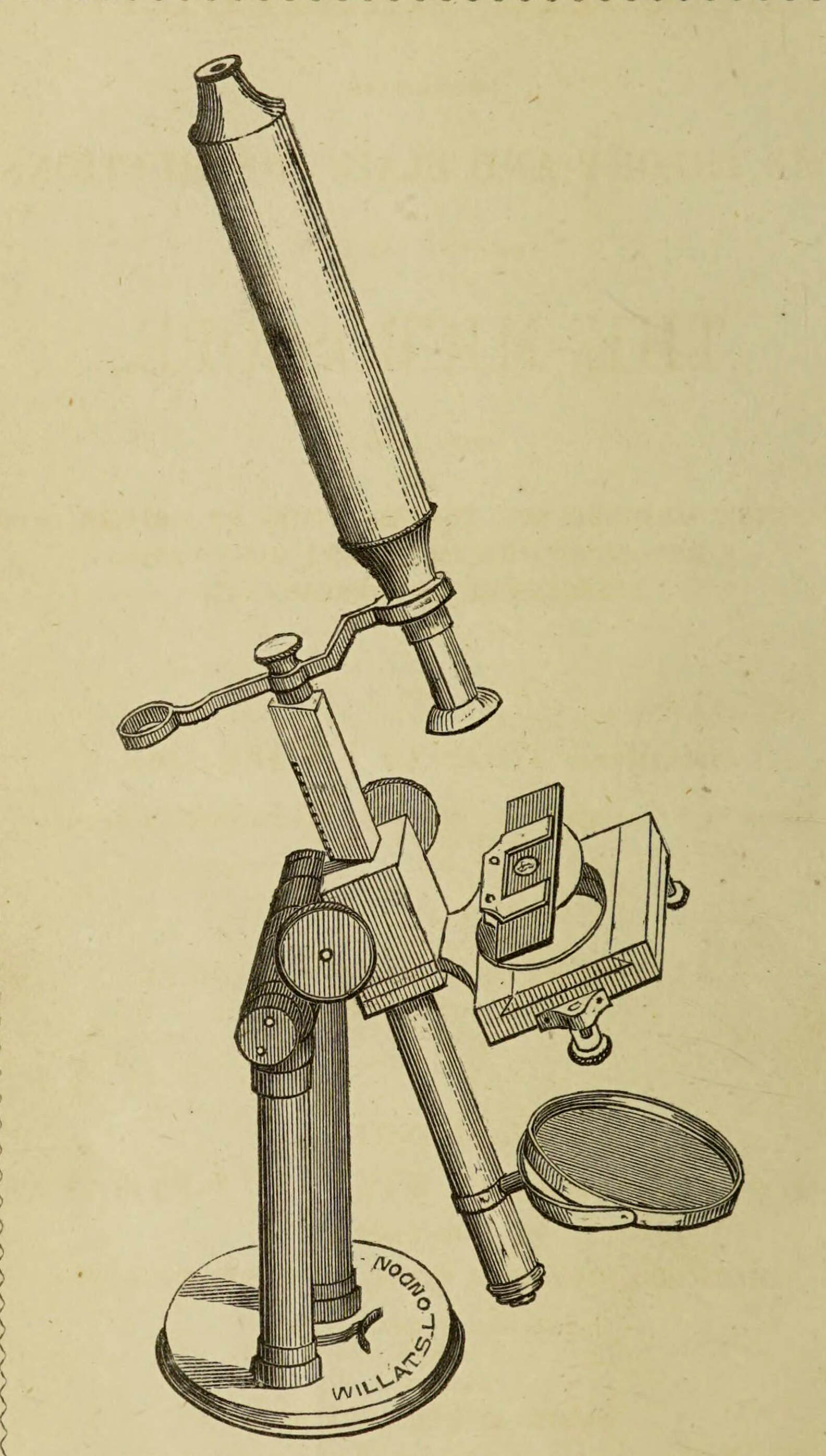
This microscope is of unmistakeably English design,and other examples exist, which are signed by different London retailers. The frontispiece to "Microscopic Manipulation containing the theory and plain instructions for the use of the Microscope" by George Thomas Fisher (Jun)and published in 1846 depicts this same
model microscope as retailed by "Willats, London". Gloria Clifton lists Thomas and Richard Willats as Opticians working between 1845-1853 from their premises at 98 Cheapside, London. They mostly sold barometers, thermometers, and electrostatic apparatus, so it is likely, that any microscopes they sold were obtained from "the trade".

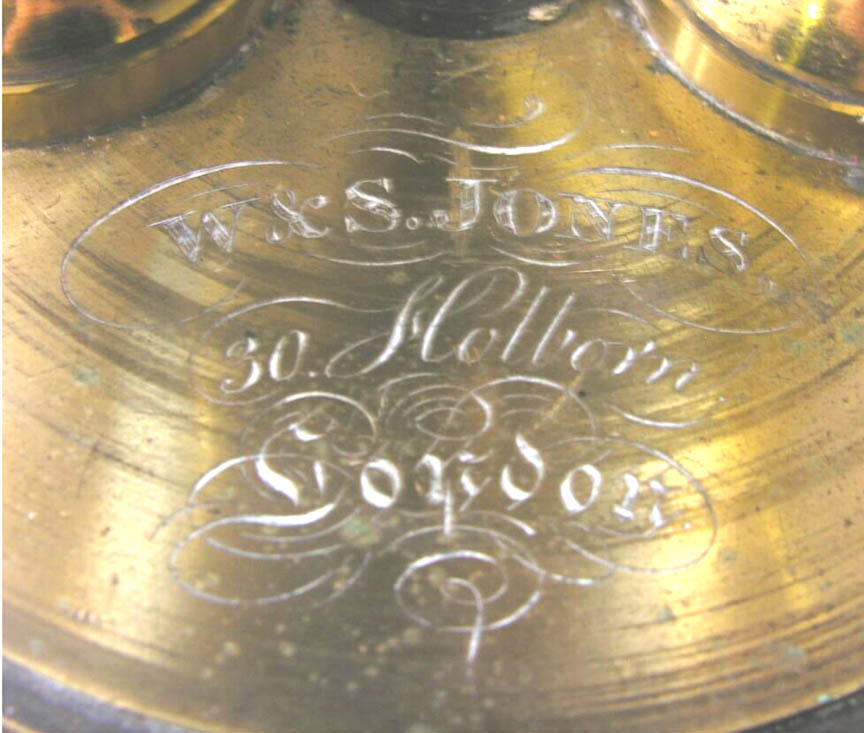
An example exists signed:W&S Jones, 30 Holborn, London
, with very similar features, but with a side-lever nosepiece adjustment (as used by Ross on early Pritchard-type models)and a mechanical stage. Samuel Jones continued the firm after his brother William's death in 1831, and traded from the 30 Holborn address from 1852 until his own death in 1859.
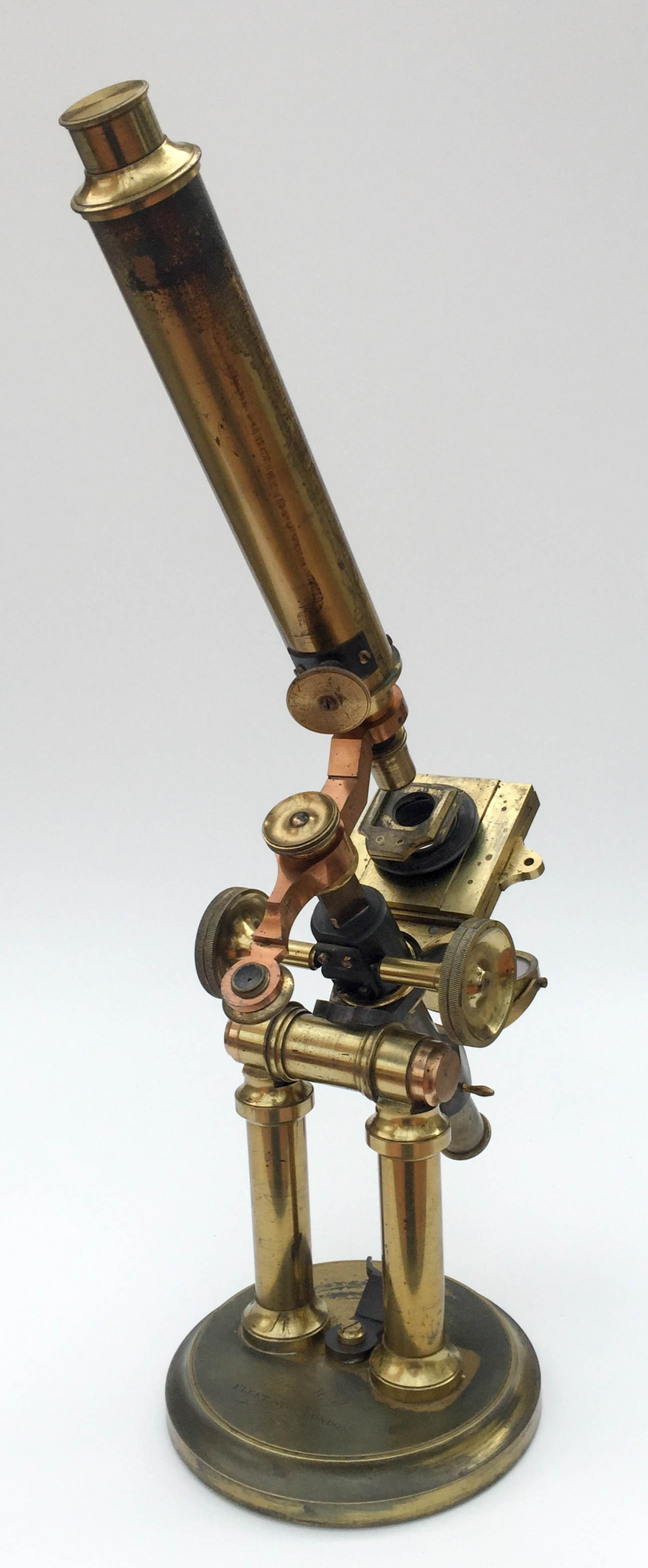
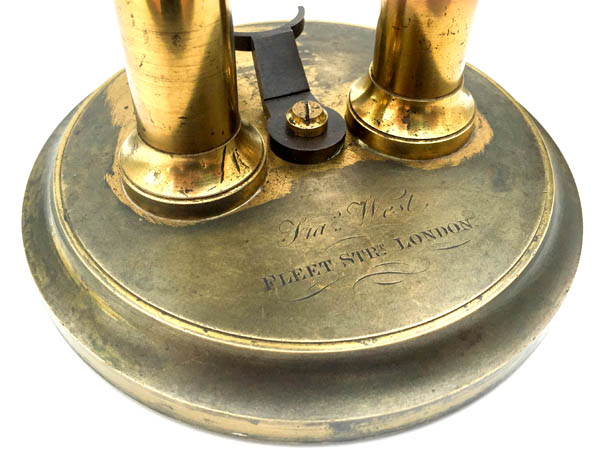 This example is signed by Francis West and features a complete double arm for use as both a dissecting and compound microscope, with a cone-type fine adjustment acting on the sprung nose-piece, and placed at the lower end of the body tube (as in a number of later Pritchard-type microscopes). There is a mechanical stage which carries the same spring stage as in the illustation of the Willats instrument. Francis West traded from 92 & 93 Fleet street
from 1849-1852, while his son, Francis Linsell set up business in 39 Southampton street,Strand, in 1849, so this microscope appears to have been retailed by the father between these dates.
This example is signed by Francis West and features a complete double arm for use as both a dissecting and compound microscope, with a cone-type fine adjustment acting on the sprung nose-piece, and placed at the lower end of the body tube (as in a number of later Pritchard-type microscopes). There is a mechanical stage which carries the same spring stage as in the illustation of the Willats instrument. Francis West traded from 92 & 93 Fleet street
from 1849-1852, while his son, Francis Linsell set up business in 39 Southampton street,Strand, in 1849, so this microscope appears to have been retailed by the father between these dates.

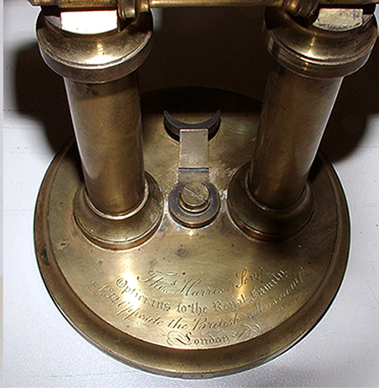 The final known example is signed:
 Thos. Harris & Son, Opticians to the Royal Family, No 52 opposite the British Museum, London
. Again, this has a double arm, mechanical stage, and superimposed spring stage, but this time, there is no fine adjustment.It would also appear, that the angled arm supporting the body tube is mounted upside down.
While it is plausible that these other examples were merely signed by retailers, the actual maker of these microscopes remains unknown. However, all appear to have a number of features in common with microscopes as sold by Andrew Pritchard: the tapered eye piece with dust cap, the (double in some cases)arm which
angles upwards towards the body tube, the horizontal lugs on the sliding mirror support, and a range of fine adjustment arrangements which can also be found on Pritchard models, while the examples as retailed by Willats and Jones have Pritchard-type objectives with integral Lieberkuhns. It is therefore possible that these microscopes all originated from a single maker associated with either Pritchard's workshop, (such as one of his own workers during the later years), or someone who made microscope stands to Pritchard's specifications. It can also be assumed that during this time, with the universal adoption of the RMS standard objective thread in 1859,nose-pieces of a number of microscopes were modified by opticians to take RMS thread objectives, at the request of owners, as is likely for this microscope. This necessitated the use of adapters for objectives with different thread. It could well be, that in the process, this particular microscope lost its double arm and a different fine adjustment was also retro-fitted. Nevertheless, this instrument represents an interesting example of the early period of brass and glass,when microscope design was still very much in a state of flux.
Text written by, and images supplied by Dr Jurriaan de Groot
We are grateful to Peter Hodds for allowing us to use his images of other examples of double-pillar microscopes.