AMPHIPLEURA PELLUCIDA ON THE DIATOM TEST SLIDES BY KLAUSE KEMP and R.I. FIRTH
AUTHOR: Barry Sobel
AMPHIPLEURA PELLUCIDA:
HISTORICAL INTRODUCTION:
 Although Amphipleura pellucida was first described in 1833 by Kützing, when it was first resolved into its alveolae is unclear. John Sloan stated he resolved the
 single frustules with daylight above or beneath the stage, with concave mirror alone in homogenous fluid or in glycerin
in an 1883 letter to the American Monthly Microscopical Journal. It is unclear to me that this meant he resolved the alveolae, the striae or neither. In any case it was Henry van Heurck who stated that he first photographed the areolae (beads
) from a silvered preparation made by Dr A.Y. Moore
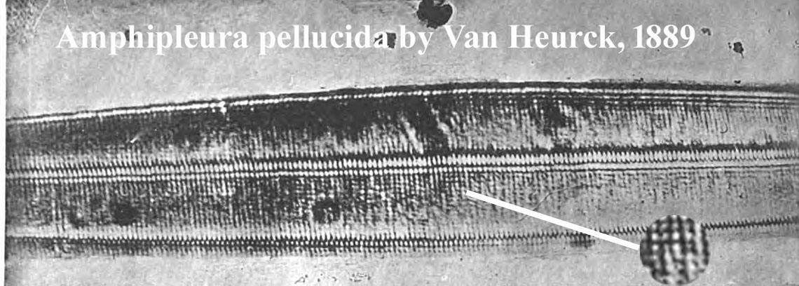
using an oil immersion Zeiss 1/12th inch objective in 1884. He stated he first published a photograph of the areole in the Bulletins de la Societe Belge de Microscopie of April 30, 1887 and I was able to find images showing resolution of the alveolae by Van Heurck in Annales de la Societe Belge De Microscopie Volume XIII of 1889 on page 14. The caption stated it was taken with a radial arm microscope, Wenham reflex condenser, and monochromatic solar lighting. Van Heurck considered this diatom to be the most difficult-to-resolve test object. Impressive images of the diatom were shown in Van Heurk's treatise The Microscope published in 1891 and later in the 1930s by Spitta in his book.
Modern Resolution:
The striae are separated by about 0.25-0.27 microns, and the areolae slightly less between 0.2 and 0.25 micron separation. The areolae are only 0.06 microns in diameter so it is amazing they can be seen at all with a light microscope. Scanning electron microscopy shows the areolae are slightly rectangular in profile, not round but because of their small size, this detail cannot be appreciated with light microscopy. For the same reason light microscopy enlargements of the areolae cannot show a sharp image. The only reason they can be visualized at all is their separation from one another, but this is only possible because this distance is just barely within the limit of the resolution of the optical microscope (0.2 microns).
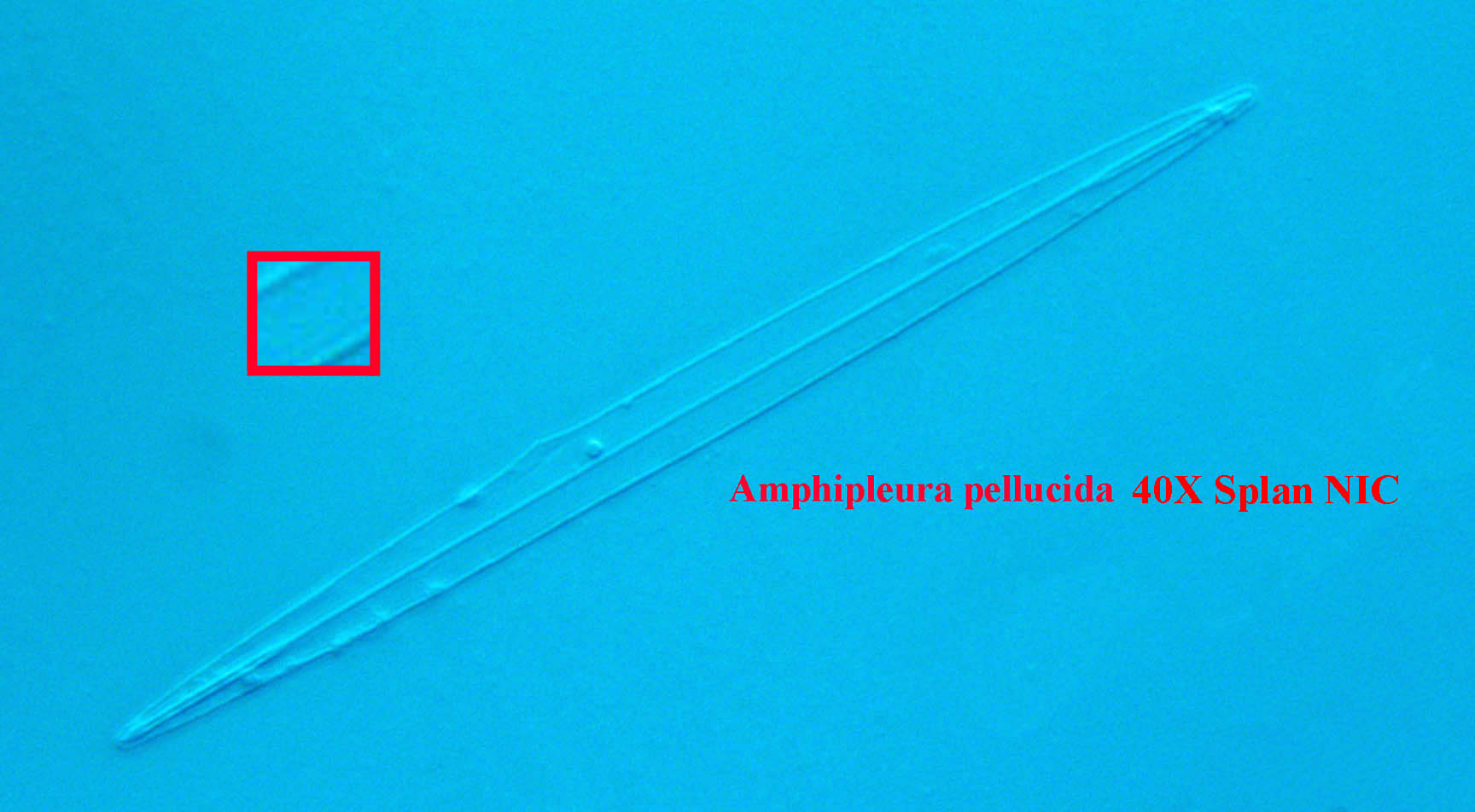
The striae of this diatom, invisible with a 40X 0.70 n.a. objective, are not easily resolved except with 100X oil immersion technique using oblique illumination or DIC. This is one of the most challenging objects for a light microscope and a specific setup has been recommended by Spitta for optimal results. Spitta stated that even without oblique lighting, high n.a. high quality objectives should show the striae though not distinctly; I could not do that with my 100X S-Plan Apo n.a. 1.35 with double oil immersion and a condenser with n.a. of 1.40. Distinct transverse striae were seen at 100X with oblique light double oil immersion.
Resolution of the areolae required oblique monochromatic green or blue light or DIC but only if mounted in Realgar* did this work well. I was unable to resolve the areolae except in a Realgar mount with any technique. The direction of the oblique light is very important in the resolution of this diatom. Spitta also pointed out that not all specimens are well-preserved, and the user should be aware of a well marked
example on the slide before proceeding. In addition, he noted that larger examples of the diatom are better than smaller ones. Barnard and Welch stated in the J.R.M.S., volume 51, pages 121-122 in 1932 that clear
resolution of the areolae would require ultraviolet oblique or dark-ground illumination.
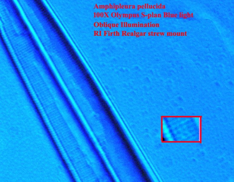
My best 100X image below was taken from a strew slide with the diatoms mounted in Realgar*, using a dichroic green filter, carefully oriented oblique lighting and crossed polarized light, using a Amscope 8MP backlit CMOS C-mount HD428N camera with 2.9u pixels and a Diagnostic Instruments C-mount adapter . It is a stack of 3 images combined with Helicon focus software. The reader should also note that the diatom chosen was partly on top of others (not shown here), meaning it would be closer to the coverslip than others in the mount underneath it. I should also mention that, although the striae were visible in the microscope with my setup, I could not see the areolae clearly in the microscope, requiring the enlarged image on the computer monitor to see them.
*Realgar (Arsenic sulfide), which has a refractive index of over 2.5, is very toxic to prepare and use until dry, and is therefore not used anymore. It also deteriorates with repeated exposure to light, so these mounts tend to deteriorate over time. I have noticed that antique slides with the specimens mounted in Realgar, seem to command a higher price than otherwise similar slides.
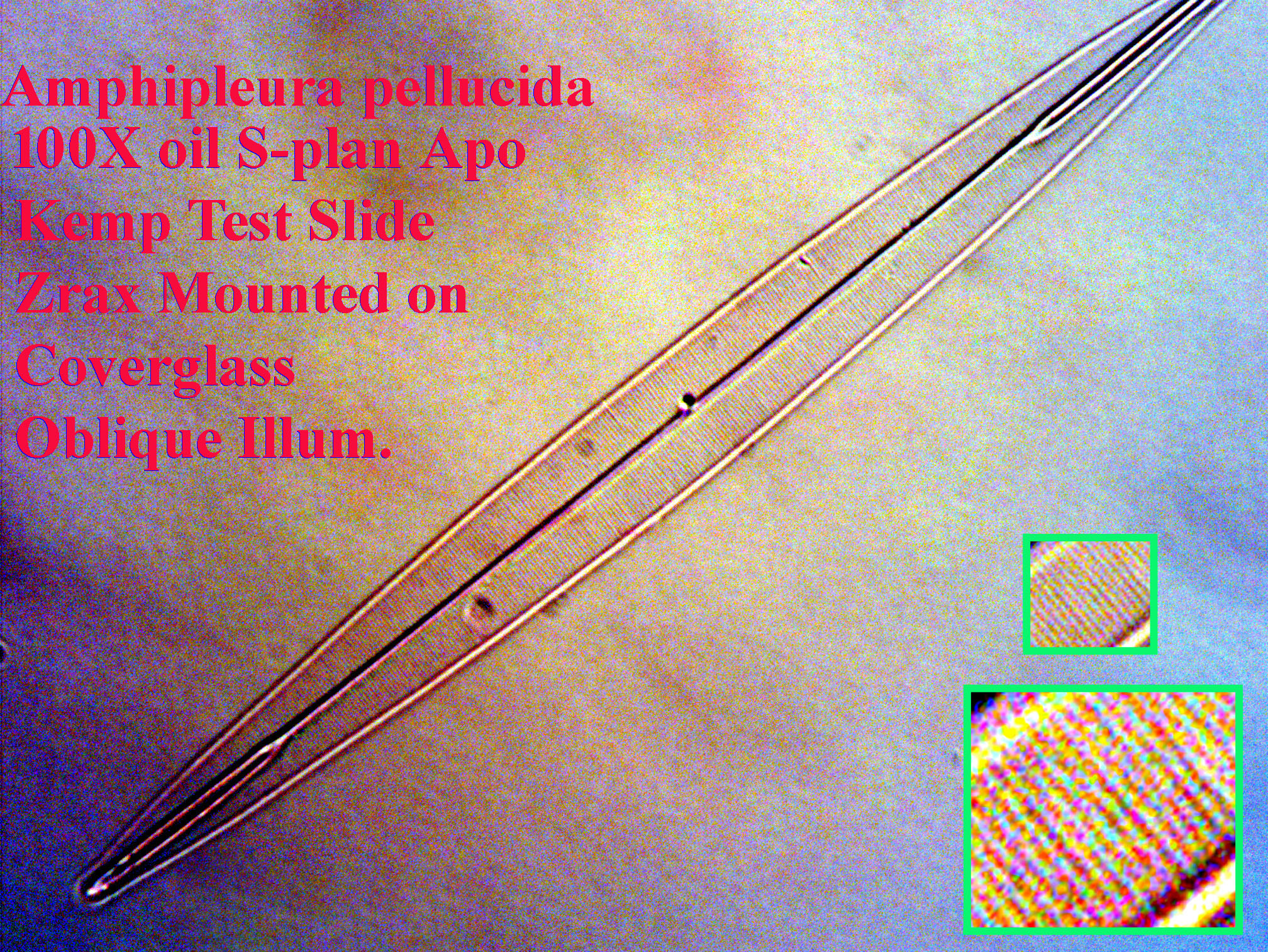
The following image is a stack of 3 images taken with a 100x Olympus S-Plan Apo objective with a n.a. of 1.35, using oblique lighting with crossed polarized light and double oil immersion; n.a. of condenser is 1.40:
.
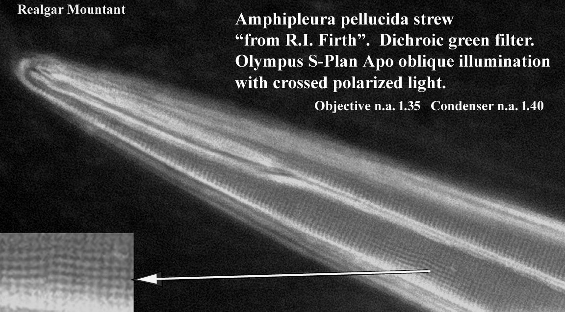
Visualizing the striae of A. pellucida, requires mounting with a high refractive index mountant like Zrax. On the other hand, areolae require, in addition, a mountant with refractive Index of more than 2.0, preferably mounted to the underside of the coverslip. Adding crossed polarized light may improve resolution slightly according to some authors. As can be seen in the above images, with Zrax, the striae can be seen with oblique illumination with oblique green or blue light provided that the source of the oblique light is oriented 90 degrees to the direction of the striae; i.e. with the long axis of the diatom oriented north-south, the light should come the northerly or southerly direction. According to Spitta (1920), the puncta are best seen with oblique light coming from a slightly different direction(e.g. southeast). But with that Zrax mount, and even with the diatom mounted on the coverslip, I have never seen the areolae well with oblique illumination, dark-ground illumination or DIC with my 100X S-Plan Apo objective and despite double oil immersion, even following Spitta's suggestions exactly. It seems the refractive index of Zrax mountant, with a R.I. of about 1.7 is just not high enough. Finally, after obtaining a Realgar strew slide, I obtained my best image using oblique dichroic green lighting with crossed polarized light using my S-Plan Apo 100x, 1.35 n.a. objective and my Olympus condenser rated at 1.40 n.a. using a Realgar mount and selecting a diatom higher up (closer to the coverslip) in the mountant than some of the others.
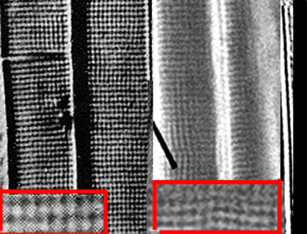
At first I thought my image was far inferior to that of Spitta, using my Amscope 8MP HDMI BSI HD428N camera, but after converting both images to the same scale, and enlarging a section of each, it became apparent that my resolution was not that far off from that of Spitta's (left side of image). In fact it was likely my specimen, on the right side of the image, was not very inferior as the parts of the diatom that are in focus in my image, are very close in resolution to Spitta's image. My belief is that Spitta probably spent many hours searching for a slide with a A. pellucida that was well preserved and flat so that the entire specimen would be in focus at the same time.