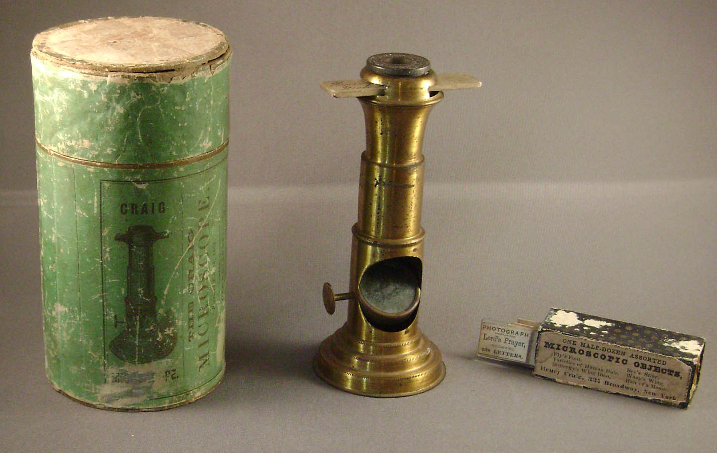
CRAIG MICROSCOPE (Lacquered Brass)
with Seven Accompanying Craig Slides including a Microphotograph
MAKER: Henry Craig
c. 1870
SIGNED ON THE LENS: 'CRAIG'S LENS, PAT'D FEBY.18.1862.'
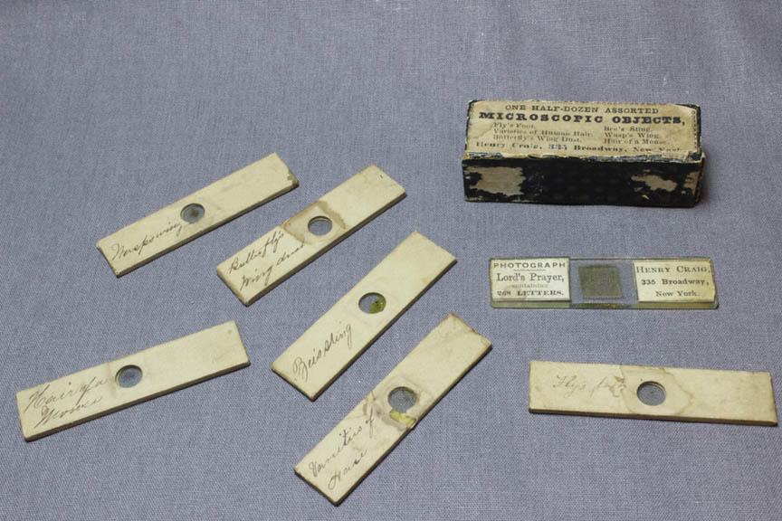
SIGNED ON THE CASE FOR SLIDES: 'ONE HALF-DOZEN ASSORTED MICROSCOPIC OBJECTS, Fly's Foot, Bee's Sting, Varieties of Human Hair, Wasp's Wing, Butterfly's Wing Dust, Hair of a Mouse,
Henry Craig, 335 Broadway, New York'
SIGNED ON THE MICROPHOTOGRAPH: 'PHOTOGRAPH Lord's Prayer, CONTAINING 268 letters, Henry Craig, 335 Broadway, New York'
Please Click On Any Picture for a Larger Version
DESCRIPTION:
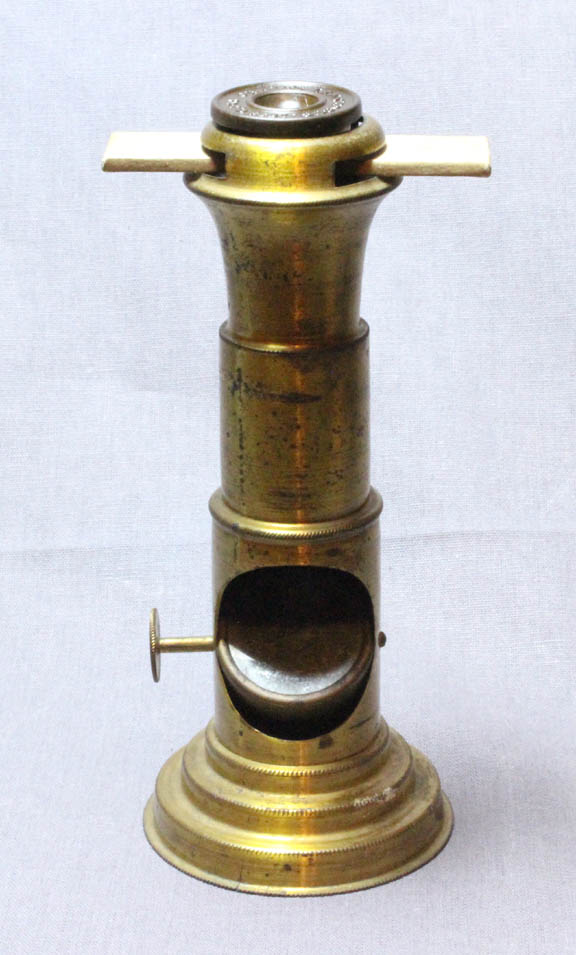
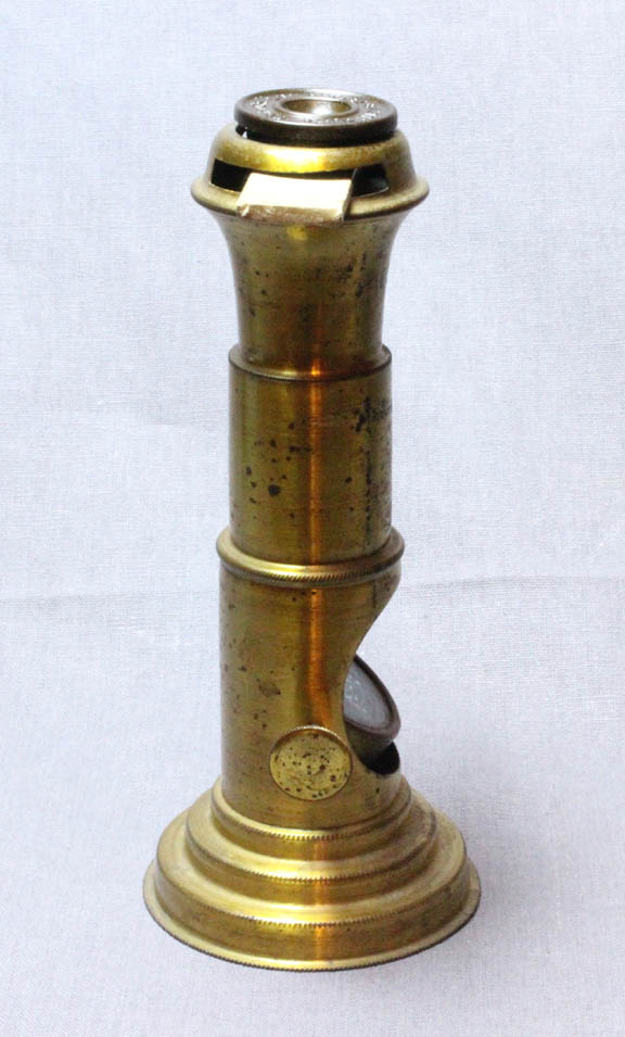
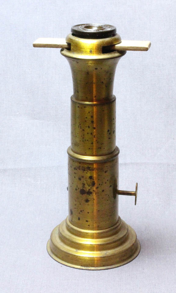
This is a simple microscope made of thin lacquered brass with a gutta-percha disc holding the lens. The lens housing is about 7/8 inches in diameter. The brass base is about 2 inches in diameter. The microscope stands about 5 inches high. The base is not weighted. There is a slot near the top to admit a slide. The mirror is controlled by a single knurled knob on one side. Inside the optical tube is a blackened disc with a single central hole to limit the size of the lightbeam entering the specimen slide.
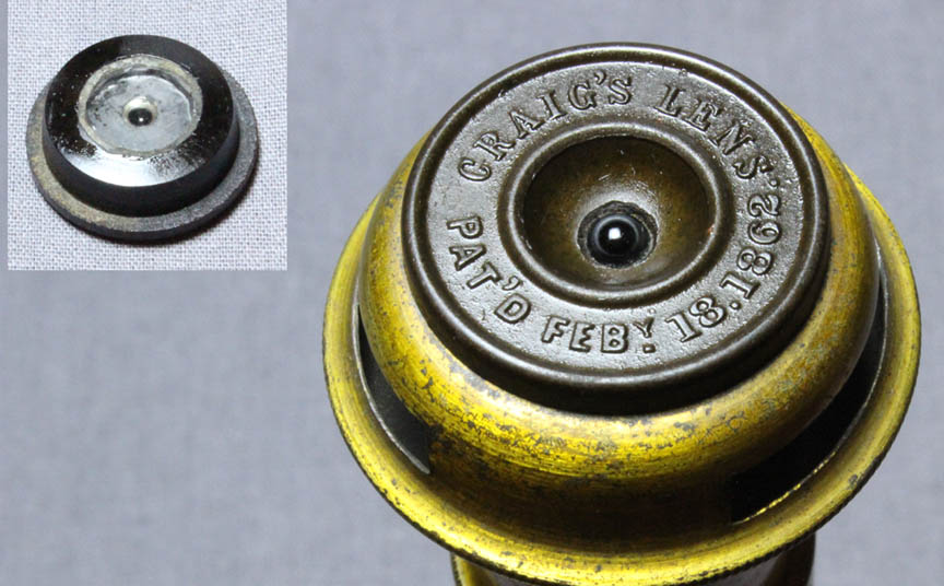
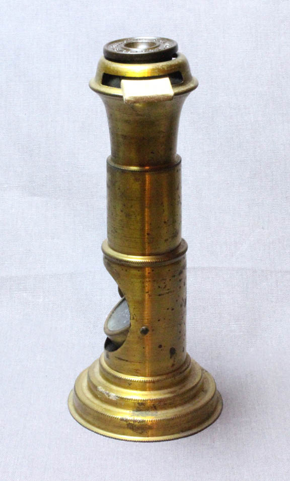
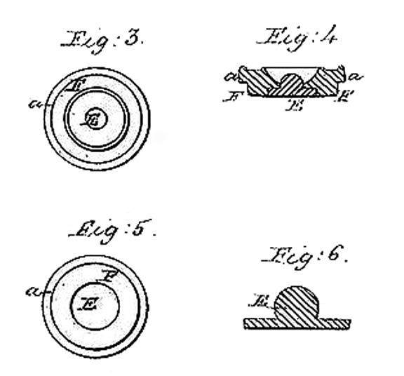 In use with a slide, the lens housing(left) is lifted up and the slide put through the slots, then the lens housing is lowered so the bottom rests on the slide. As shown in the diagrams from the patent(right), the lens is actually composed of a sphere of flint glass, fused to a flat piece of crown glass, thus making it somewhat achromatic. The lens bottom is covered with a thin piece of glass at the focal point, such that objects in contact with it would be in focus; it was also intended to have specimens placed directly on this bottom surface, adhering to it by surface tension, as would a drop of pond water. The focus is fixed at the bottom of the lens housing, so it is impossible to do fine work, especially with a specimen mounted under a coverslip. Nevertheless it does allow one to read the words of the microphotograph, and have fairly good views of the specimens in the slide set, which, with thin coverslips, are in reasonable focus. After cleaning, the lens in this example is still not perfect with a thin layer of debris or imperfections that I could not remove.
The cylindrical cardboard case bears the distributer's name:
C.H. WHEELER & CO.,Nos 5 & 7 Essex Street Boston,SOLE AGENTS FOR THE NEW ENGLAND STATES
The green cylindrical box for the microscope is made of cardboard and, like many examples, has lost one of the two thin ends. It gives instructions on how to view drops of liquid containing subjects by applying them to the bottom of the lens and then reversing the lens on the microscope to its usual position, to view the contents of the drop. It also explains that thin coverslips must be used with the usual objects, and that slides prepared with thin slips like this can be obtained from the sellers of this microscope.
The enclosed slides are 2 1/4 x 1/2 inch and have thin coverslips under their paper covers. The original box for the slides is about 7/8 inches thick on each side. An inner, originally 5 sided, box slides into the outer box, but its ends are missing. This box of six slides, actually containing seven with the extra microphotograph, even has room for one or two more slides. Not only did Craig offer a six slide set, but also a 12 slide set in a wider box.
HISTORY OF THE CRAIG MICROSCOPE AND SLIDES
This microscope was patented in 1862 and apparently produced for about 10 years. These flimsy microscopes from over 150 years ago, are relatively uncommon in good condition when complete with the lens; the box and slides are even less common. They originally sold without slides for $2 then, about $30 today. This is quite expensive for what the buyer was paying for, and for the same price a much better magnifier or simple microscope could have been purchased. In fact, it is almost easier to use the Craig lens directly on the slide without the rest of the microscope. Furthermore, opaque objects cannot be examined with this lens designed for contact focusing.
A similar microscope, of very slightly different construction was the Globe Microscope
made from about 1872 to 1878. An example of the Globe microscope is on this site.
For a detailed history of Craig microscopes, including its other variations in gutta percha and, in the last years of production, nickel plated, and the somewhat similar Globe microscope, please see the article about them on Microscopist.net.
A ROUGH TIMELINE OF THE CRAIG AND GLOBE MICROSCOPES:
February 1862: Henry Craig was issued Patent
Early 1860s: Craig Wheeler distributed the Craig Microscope as the Sole Agent for New England
from Boston, MA
1863: John Eliss became the distributor
1864: Henry Craig Died
1864-1866: John Ellis sold (and likely made) Craig microscopes
1868: George Meade of Racine Wisconsin sold Craig Microscopes via a PO Box in Chicago, Ill
1870-1871: Theodore Tusch sold Craig microscopes from NY City
1872-1878: George Meade made
and sold the Globe Microscope, similar to the Craig Model but with a single spherical lens which could be focused. He also advertised the sale of microscope slides that could be used with this microscope.
WARNING-PLEASE NOTE: If you are intending to buy one of these for your collection, please be careful. Many are sold without the lens, and are therefore of minimal or no value; without the lens, its just a cheap thin piece of metal without any use as a microscope!