5-FORM DIATOM TEST SLIDE BY J.D.MÖLLER
c. 1st Half of 20th C.
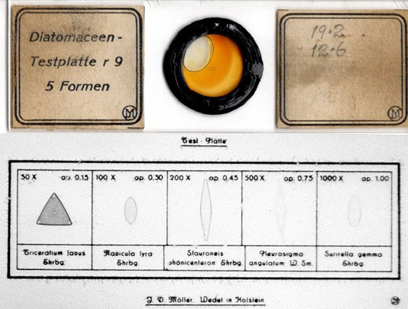
This is a diatom test slide by the Möller company. The diatoms are mounted within a microphotograph. It probably dates to the first quarter or perhaps the second quarter of the 20th century. As shown above, it is labeled:
Diatomaceen-,Testplatte r9, 5 Formen
with the J.D.Möller insignia on the lower right hand corner of the left side label. The right side label also has the insignia and has handwritten on it 19.2, 12.6
. Overall the slide is in good condition though the mountant has darkened with age, and with a bubble in the mountant (which does not enter the field of the test diatoms). Also as shown above, the title of Test-Platte
sits above, and J.D. Möller Wedel in Holstein
sits below grid enclosing the diatoms. For a detailed history of Möller and his company, please see Brian Stevenson's review on microscopist.net
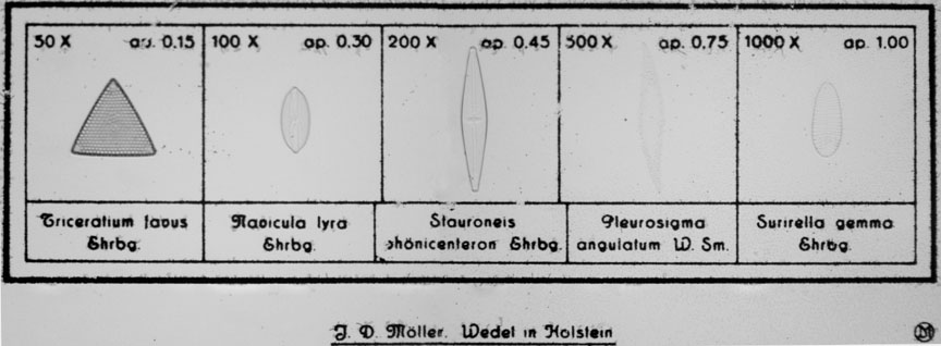
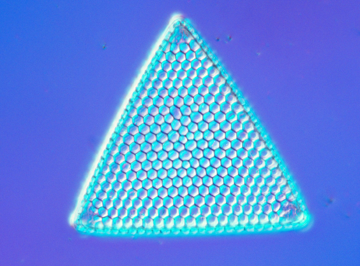
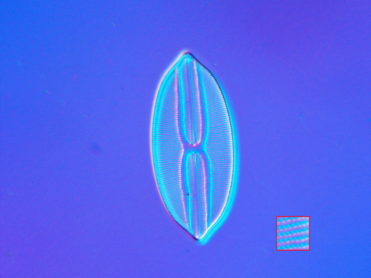
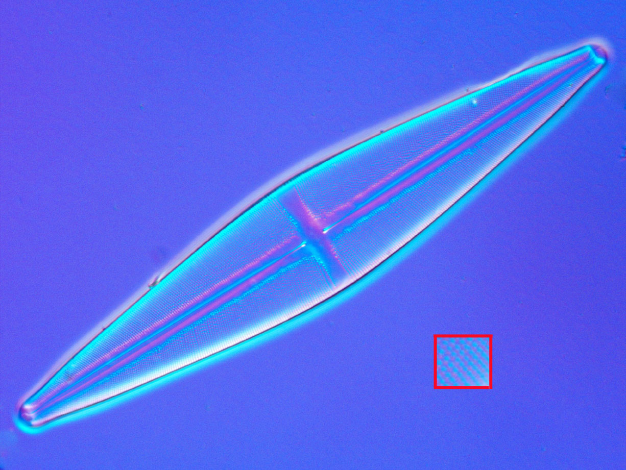
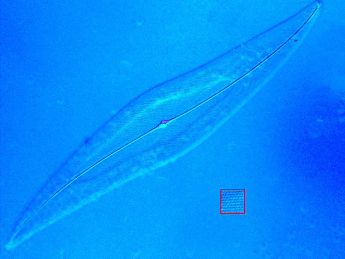
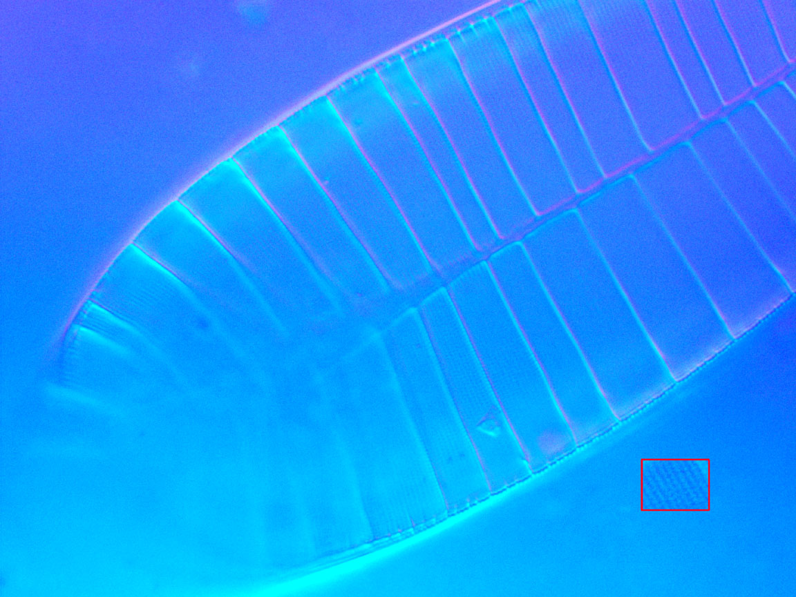
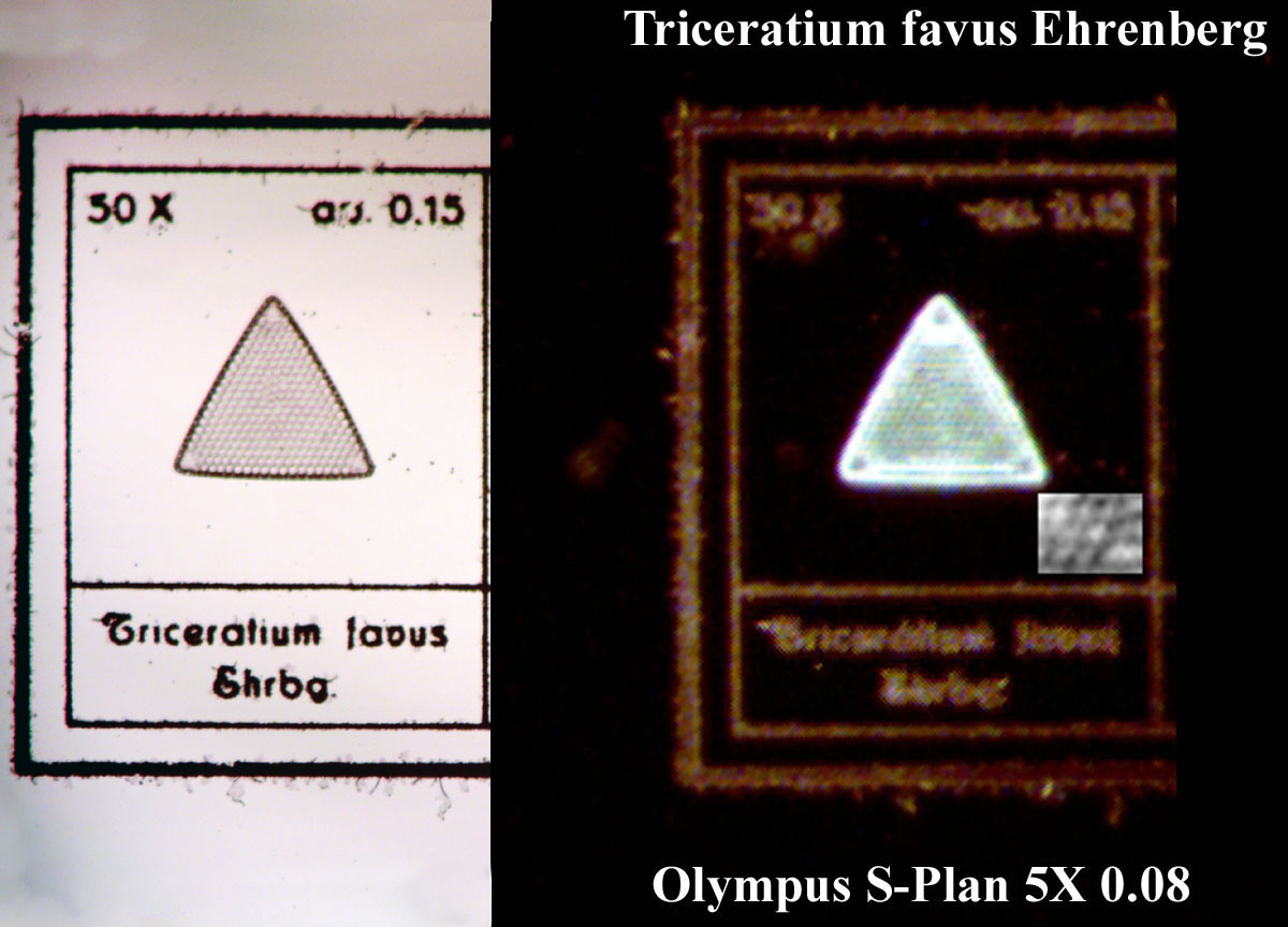
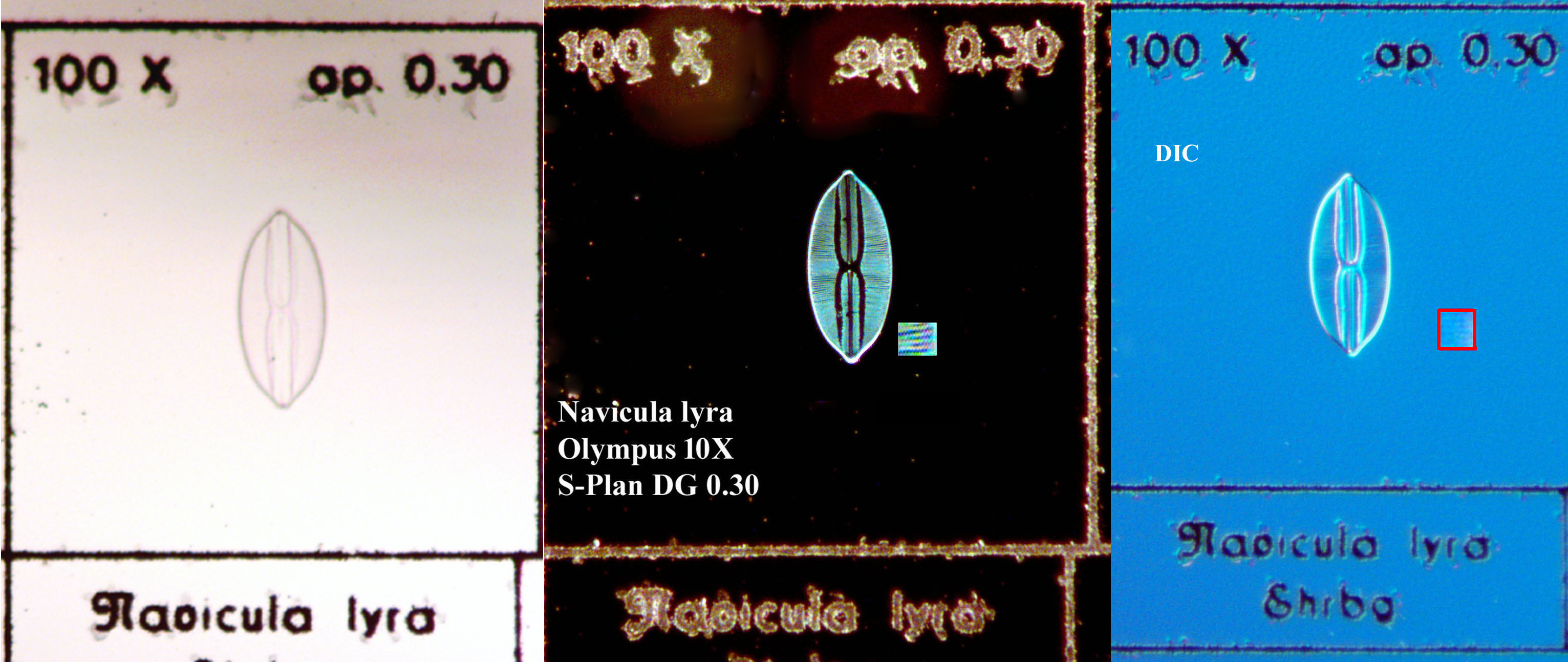
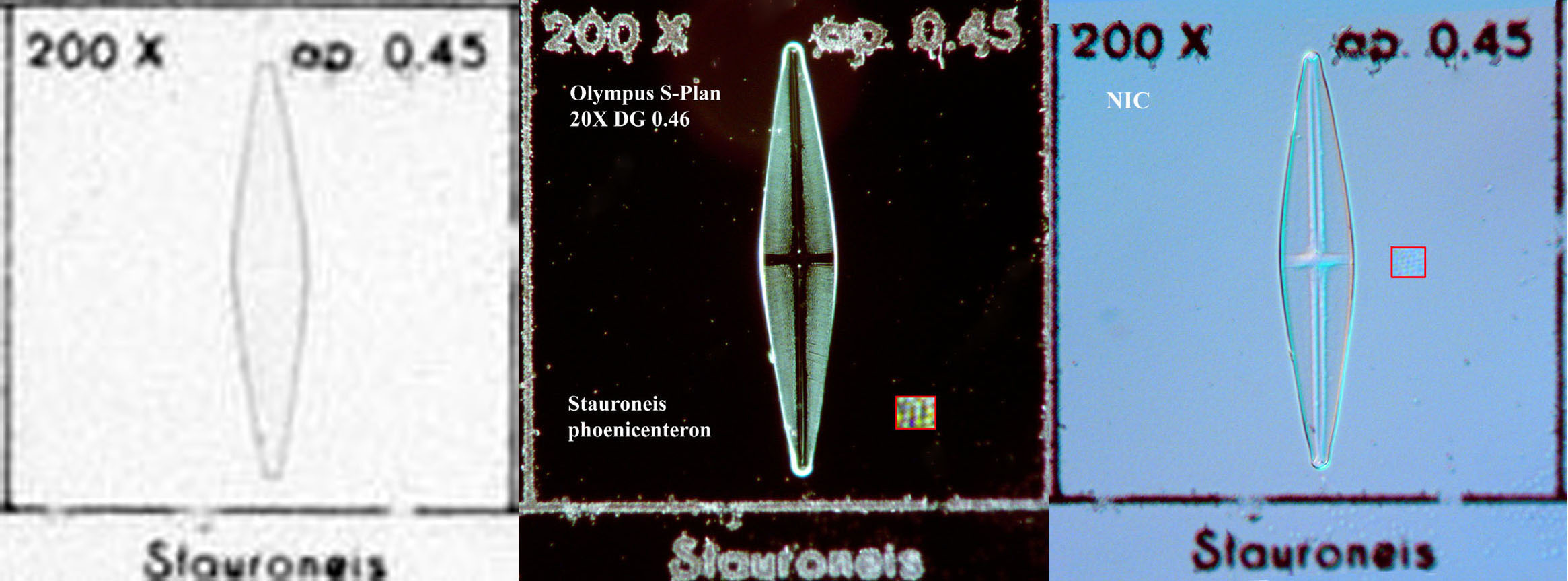
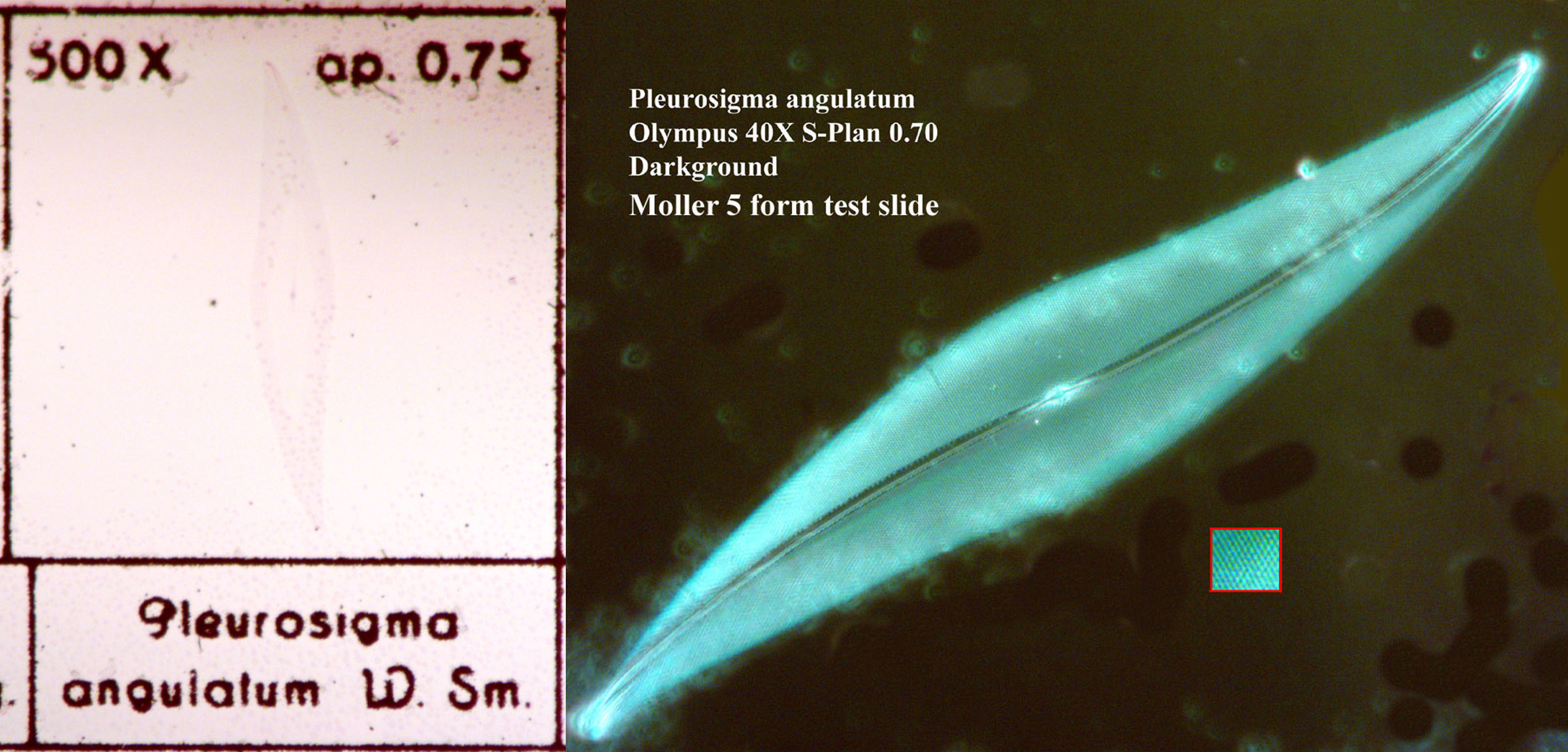
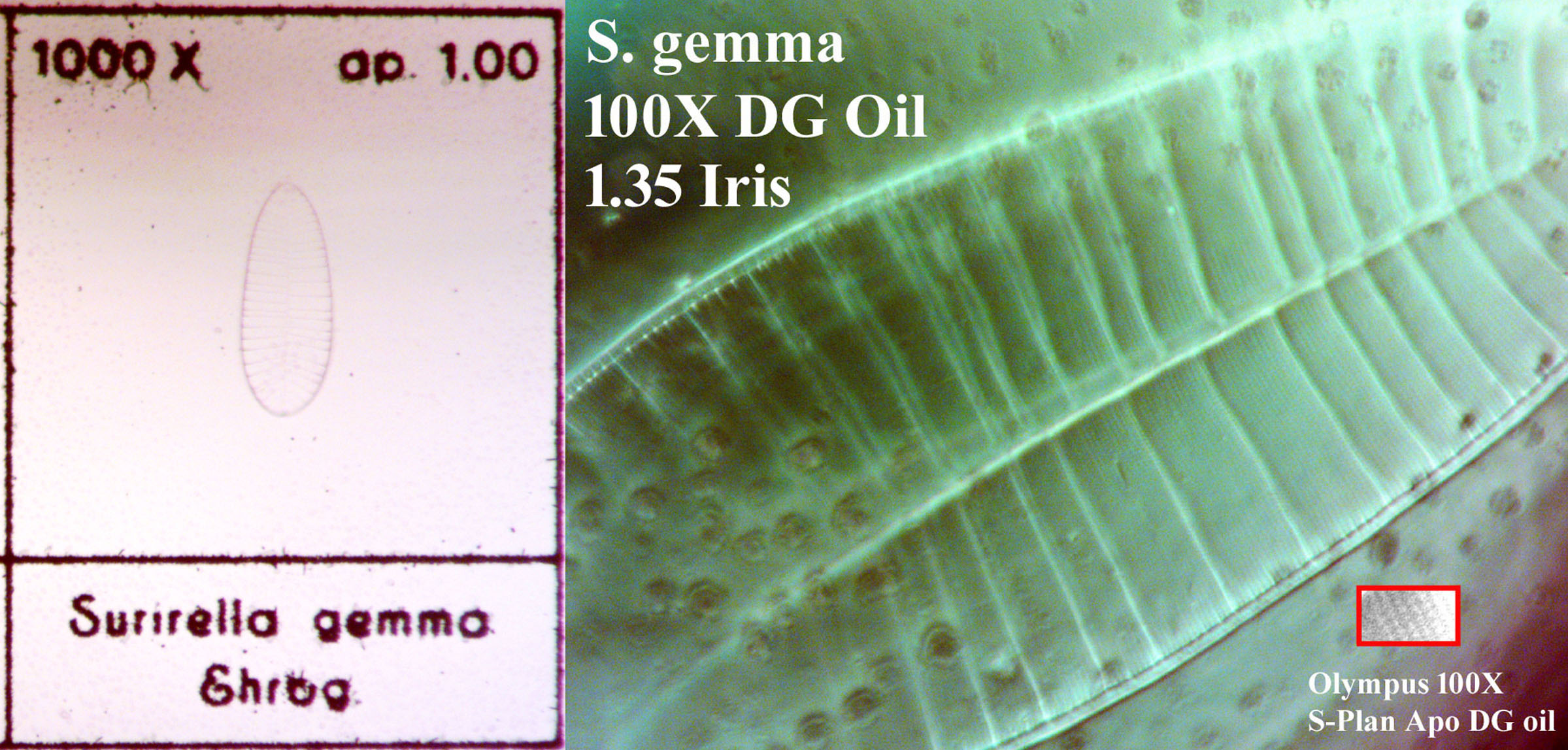
The slide contains 5 diatoms, each in its own cell within a photographic grid. The diatoms are arranged requiring higher resolution and magnification to be resolved down to the puncta as one progresses from left to right. The suggested requirements for each are noted in the cell of each diatom. These particular diatoms were well known for being used as test objects when this slide was made, and are sometimes still used for this purpose more recently and even today, though the exact diatoms used as test objects vary a bit from mounter to mounter. From left to right the diatoms are Triceratium favus, Navicula lyra, Stauroneis phoenicenteron, Pleurosigma angulatum, and Surirella gemma. Using my Vanox microscope, I was able to determine that simple brightfield Kohler illumination could not resolve these diatoms down to the puncta except for Triceratium. Although both DIC illumination and darkground illumination worked, darkground gave slightly better resolution. Oblique illumination was only marginally effective for all the diatoms except for pleurosigma which was well resolved by it. Darkground illumination was the most consistent method to resolve each diatom down to the puncta using the objectives noted, as shown in each of the following images. The insets are from the same images, but enlarged to show the puncta more clearly. Unfortunately in dark ground illumination, the printed information does not image well where brightfield imaging is superior. The dry darkground condenser has a n.a. of up to 0.86 and the oil immersion darkground condenser has a n.a. of about 1.35 when used with the 100X S-Plan Apo objective. I found that the suggested objectives for Navicula lyra and Stauroneis phoenicenteron were the absolute minimum required resolving the striae well but the puncta just barely. I found this to be true with other slides containing these same diatoms by other preparers as well.
Below are shown the each of these diatoms in DIC, the first four with a 40X objective, the last with 100X, illustrating the beauty of these specimens: