PLEUROSIGMA ANGULATUM:
This is a famous diatom used for many years as a standard test object. The genus Pleurosigma differs from Gyrosigma in that the areolae of the latter form striae only in two directions. Pleurosigma is characterized by areolae which can be traced longitudinally, transversely and diagonally.
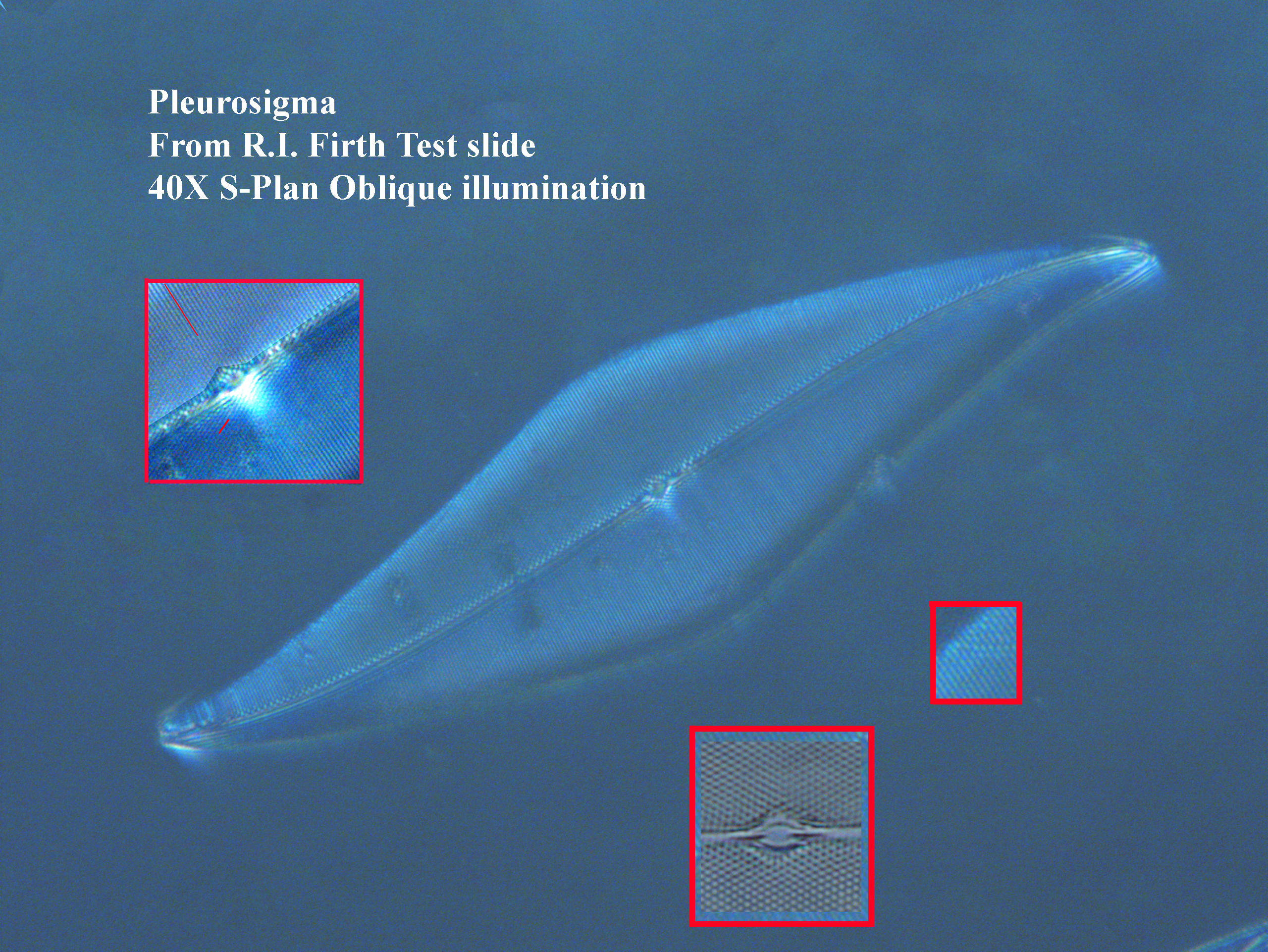
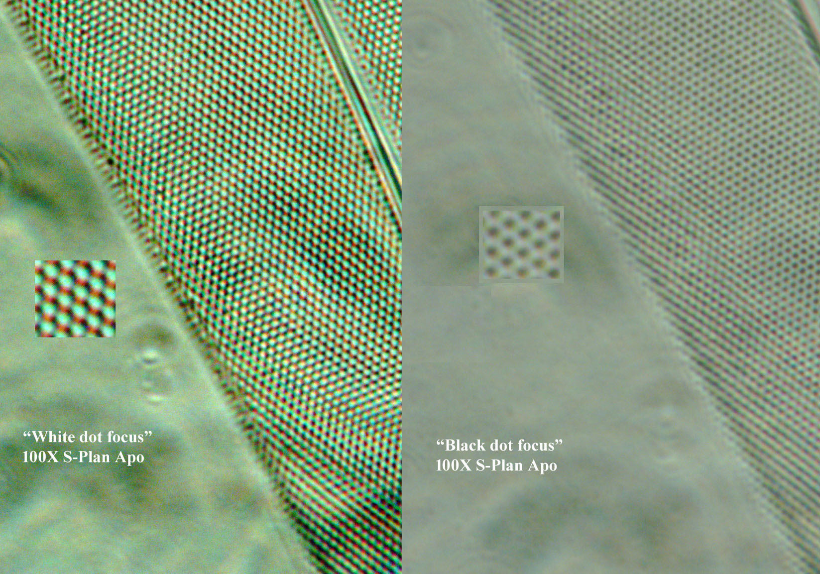
As one uses objectives with higher n.a., increasing resolution, one notes that one can focus on the areolae and get a black dot
effect or a white dot effect.
This effect was used, in prior times, by comparing the appearance each way, to judge the quality of high power objectives. This black and white effect is likely due to optical effects at different distances of focus, not a structural difference. The black dot effect is seen with deeper focus, the white is seen with focusing on the surface of the diatom.
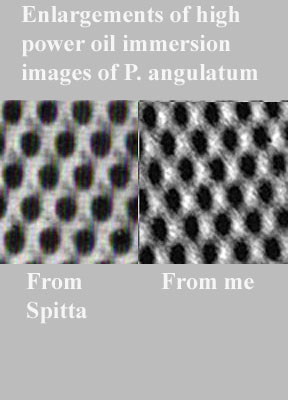
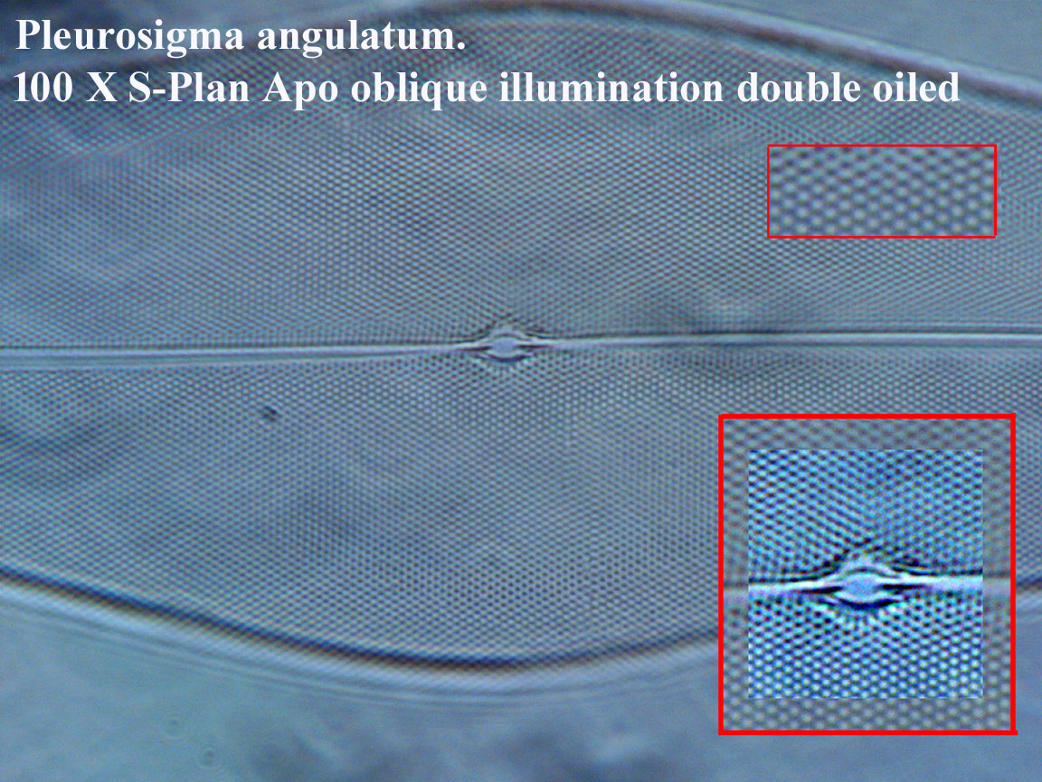
Another interesting effect is that when using a light microscope, with maximum resolution (as shown in the final images on this page), there seems to be a vague suggestion of a hexagonal chamber to the areolae. This turns out to be an artifact of the diffraction of light as it passes through these tiny areolae because, as shown by electron microscopy, the areolae do not actually have hexagonal walls. Although the spacing between the areolae is within the limits of separation of the light microscope, the resolution of a light microscope cannot reveal the true shape of the areolae accurately, as the size of individual areolae means their details must be beyond the limit of light microscope resolution.
In fact, the structure of the areolae are quite complex. In Scanning Electron Microscope images, each valve of Pleurosigma species turns out to be a sandwich. There is an internal layer with roundish pores that are closed off by a thin sieve-membrane (called a hymen), and an external layer with slit-like openings. Both openings are slightly oval in shape; they are not round nor hexagonal. Nevertheless, the apparent hexagonal shape seen with high power light microscopy is interesting to see.
P. angulatum can be used to illustrate several principals of microscopy. One of these is the principal of empty magnification
. This is illustrated by the first two images below. When used as a test object, the striae of P. angulatum can be resolved into areolae with an n.a. of about 0.52 or greater. Although both the first and second images are both taken with a 20X objective, only the higher n.a. objective can resolve the areolae. This diatom can also illustrate the effects of proper illumination, especially the optimal setting for the aperture diaphragm, where the image appears washed out if it is open too far reflecting loss of contrast, loss of resolution if it is closed to far, and optimal resolution with optimal contrast when closed to a bit less than the diameter of the back element. It also illustrates the advantage of oblique illumination for increasing contrast when the details are so small.
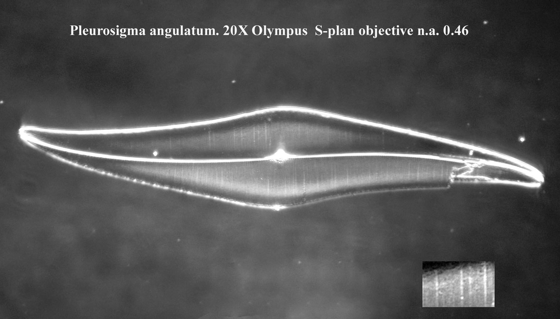
Pleurosigma angulatum 20X objective n.a. 0.46; note that the areolae are not resolved even with oblique lighting:
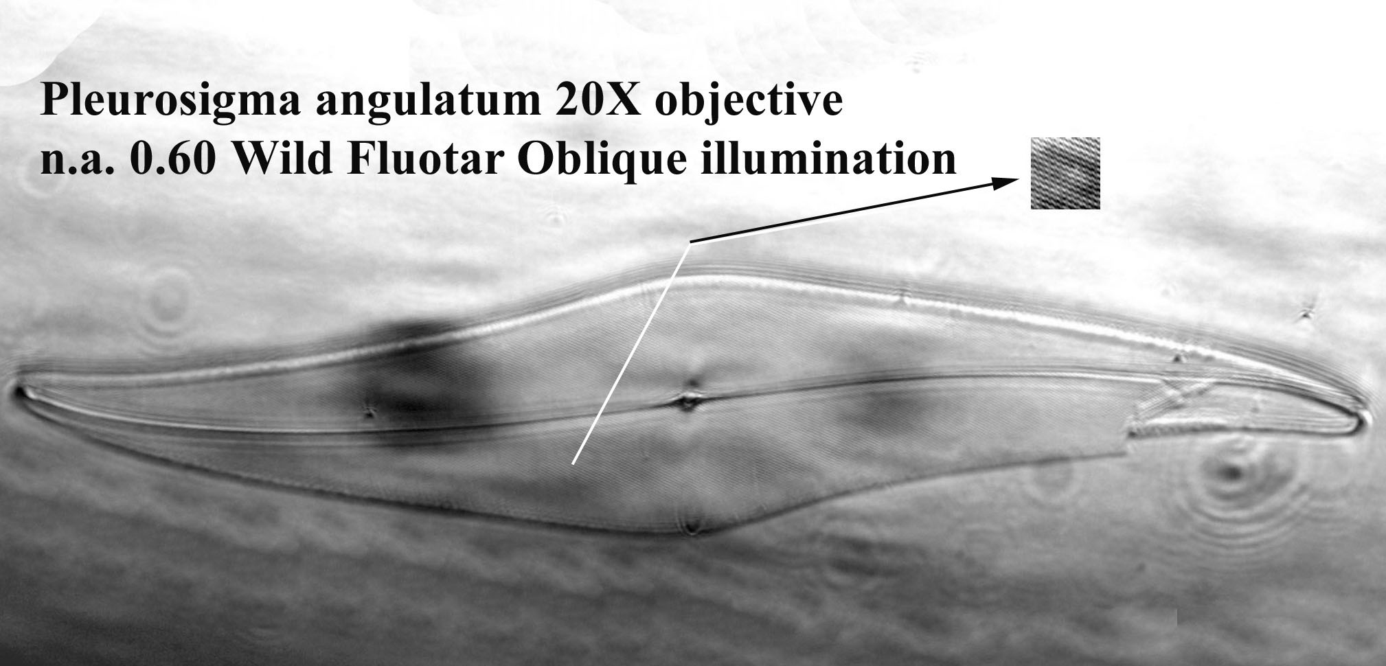
Pleurosigma angulatum 20X objective n.a. 0.60; note that, despite the same magnification, the areolae are now resolved:
Pleurosigma angulatum 40X objective brightfield:
Pleurosigma angulatum 40X objective Oblique Illumination:
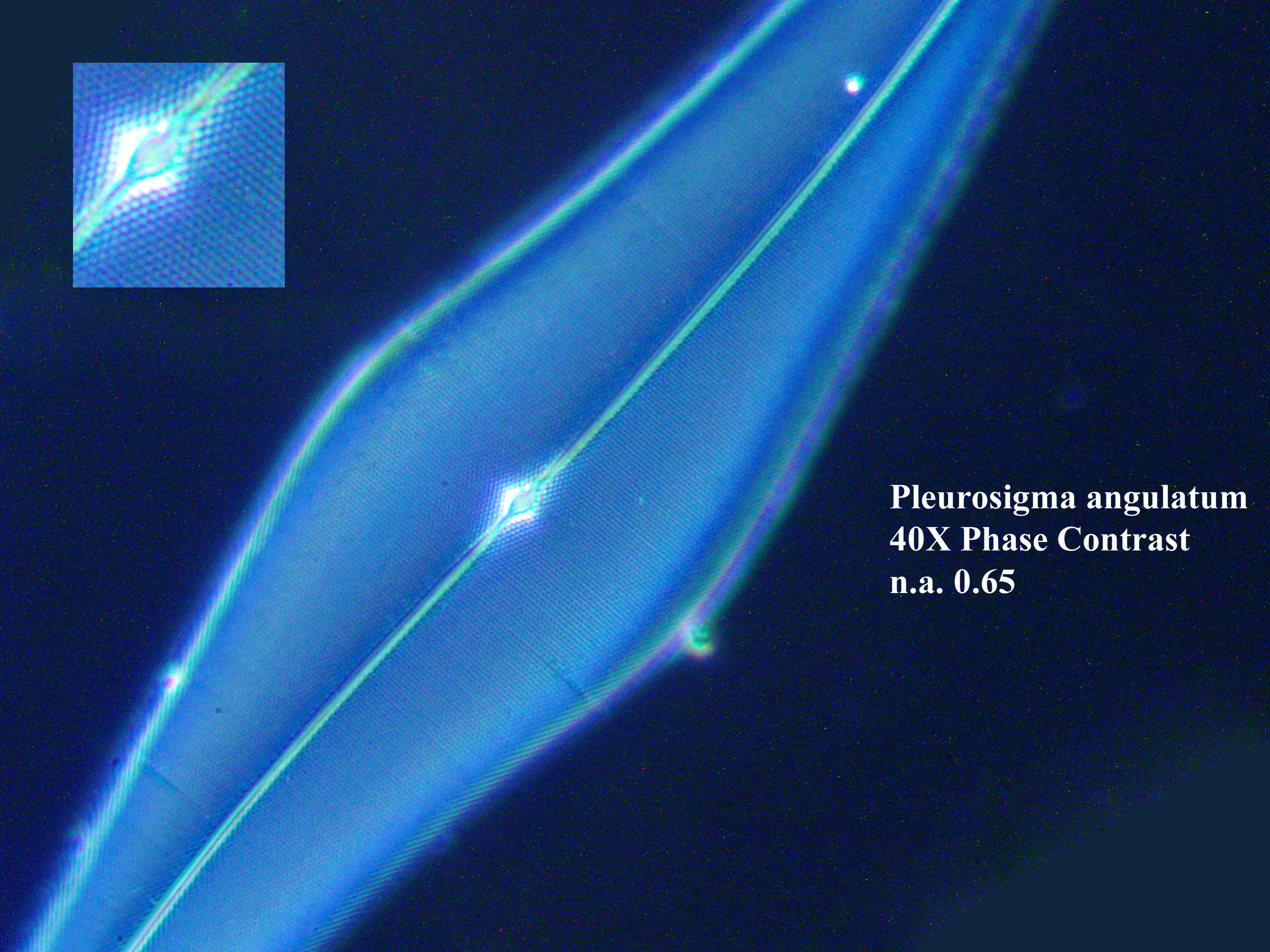
Pleurosigma angulatum 40X n.a. 0.65 objective Phase contrast Illumination; although phase contrast reduces the effective n.a. of the objective, the high base n.a. remains high enough to resolve the areolae:
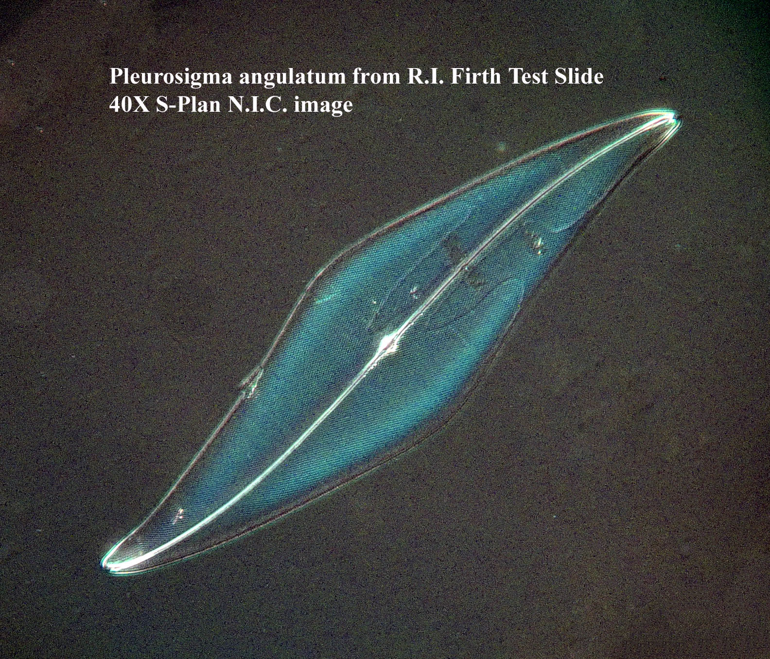
Pleurosigma angulatum 40X objective NIC:
Pleurosigma angulatum 100X S-Plan Apo objective Oblique Illumination:
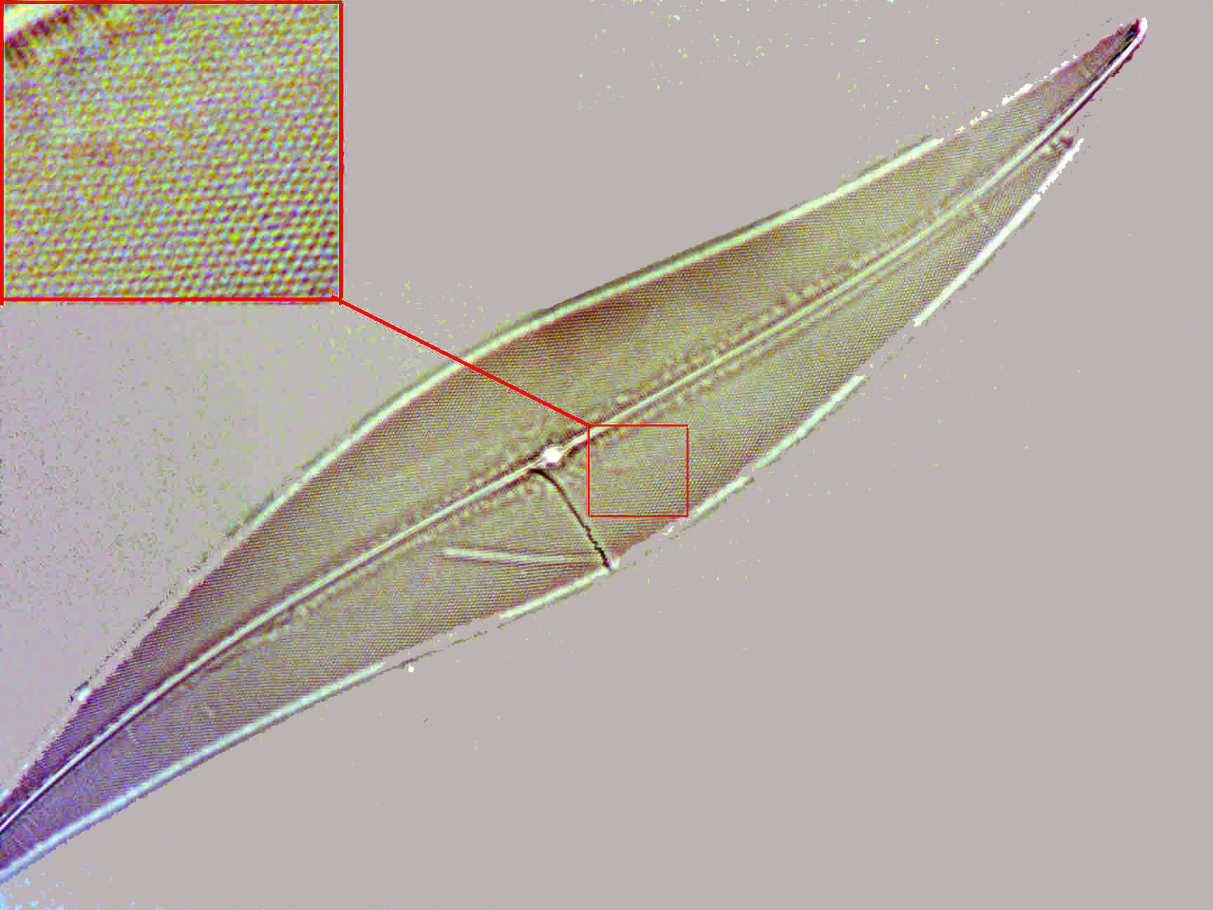
Pleurosigma angulatum at high power illustrating the white dot and black dot
effect. 100X S-Plan Apo objective Oblique Illumination:
Pleurosigma angulatum at high power illustrating the pseudo-hexagonal appearance of the areolae when viewed under high power light microscopy: