COMPOUND BINOCULAR MICROSCOPE
MAKER:Smith & Beck
MODEL:'Best No 1'
c.1858-1865
SIGNED:'Smith & Beck, 6 Coleman St, LONDON'
SERIAL NUMBER: 1873
POLARIZED LIGHT ACCESSORIES
POLARIZED LIGHT APPARATUS:
The polarized light apparatus supplied with this microscope is extensive (for the time period), and includes all the apparatus to demonstrate all the double image prism experiments described by Legg in the Transactions of the Microscopic Society of London, Vol 1 of December 1846, and also described in Richard Beck'sTreatise on the Construction, Proper Use and Capabilities of Smith, Beck, and Beck's Achromatic MicroscopesTreatise of 1865 It also included fittings for standard studies of specimens under polarized light with or without a variety of selenites. Scroll down for more information and accessories.
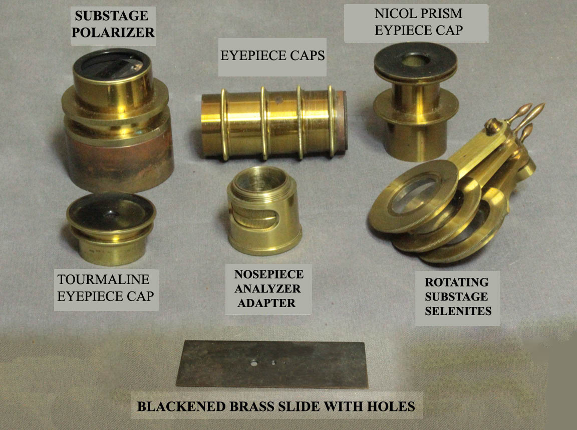
 This microscope was supplied with a large calcite prism polarizer, which mounts to the underside of the adjustable substage.
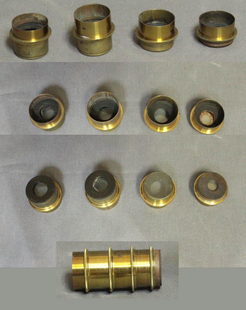
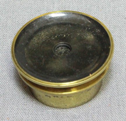
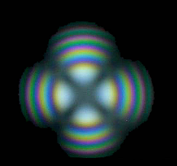 Four eyepiece caps are stored stacked on each other. These include two double image prism eyepiece caps(left two), a selenite eyepiece cap(third from left), and a crystal to illustrate conoscopic interference figures(fourth from left). One of the double image caps is slightly taller than the other. The image to the right illustrates a conoscopic figure produced with the crystal shown here. See the double image prism page for an earlier Ross microscope for illustrations of some of the experiments using double image prism.
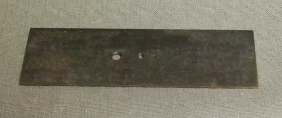 This is the blackened brass slide with three different size holes for the double image prism experiments.
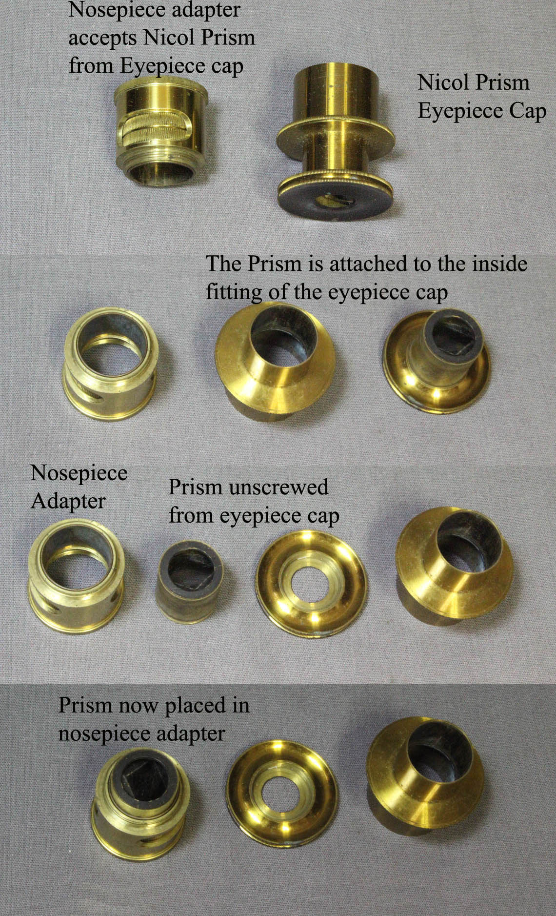
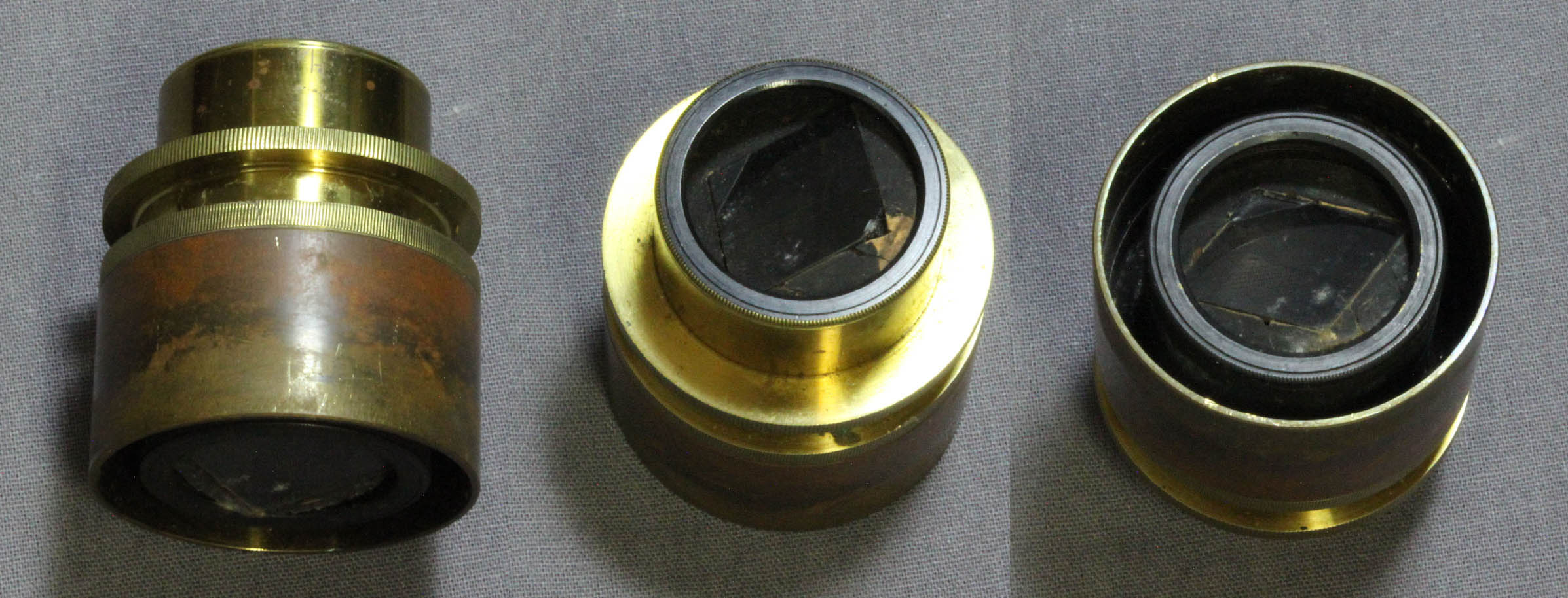
 There are two additional separate eyepiece caps which include a Nicol prism analyzer, and a tourmaline eyepiece cap. These are both used as analyzers. The Nicol calcite prism, shown here with the nosepiece adapter to the left, works well but suffers from vignetting (reduced field of view). The calcite prism analyzer eyecap has the prism assembly screwed into the bottom of the blackened eyecap, and this assembly slides inside an outer sleeve that fits over the eyepieces. After being unscrewed from the eyecap adapter, the analyzer prism fits into the nosepiece adapter, which reduces vignetting compared to using it as a eyecap analyzer. But vignetting and distortion still occur if this nosepiece analyzer is used with the binocular tubes. In later years, James Swift devised a sliding analyzer positioned above the Wenham prism, to allow use with binocular tubes without as much distortion.
The tourmaline eyecap(right) has a larger field of view than the analyzer eyecap, but imparts color artifact to the specimen. The tourmaline eyecap is labeled in white script tourmaline.
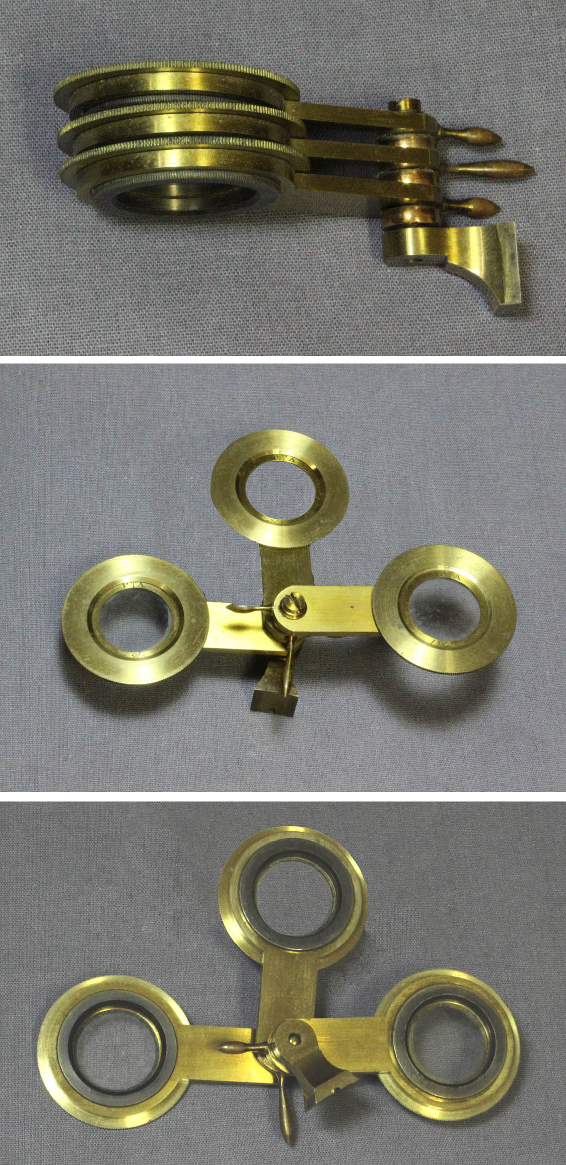
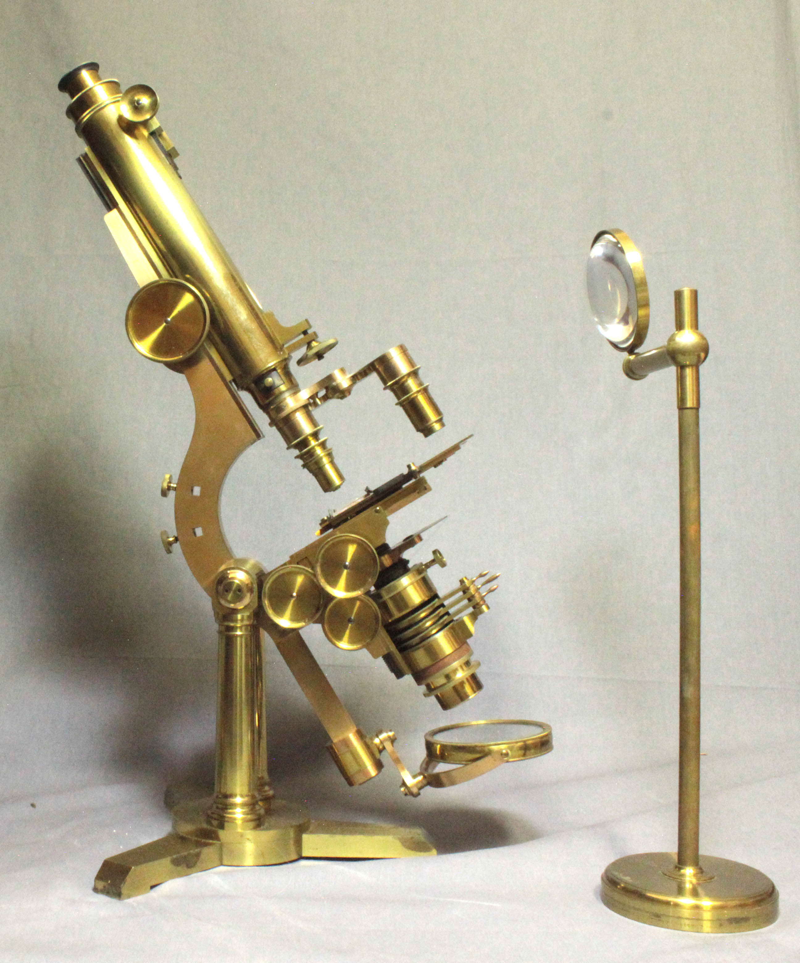 A set of three stacked selenites fits into the substage assembly via a dovetail. Each selenite can be placed in or taken out of the optical axis via a small lever handle. Each selenite can be individually rotated. This allows a large number of different color combinations. The image to the right shows the microscope set up for polarized light work with the selenites installed on the substage with the polarizer below and condenser above.