test-objectsin my collection along with associated information. It is important to realize that modern mounts of these same objects may differ from the ones originally described, and therefore current conclusions may not be the same as those in years gone by. In some cases, resolution is better than in the past using current equipment, but in some cases this is not true due to factors such as the type of mounting material, thickness of coverslip, thickness of slide and if the subject is mounted on the slide or the bottom of the coverslip. If the user is interested in seriously determining the quality of an objective, the objective tests such as the Abbe test plate, an Apertometer, etc may be best. If interested in exploring the tests like they once were, then the subjective tests will be appropriate, and also entertaining. This page is about the natural test subjects.
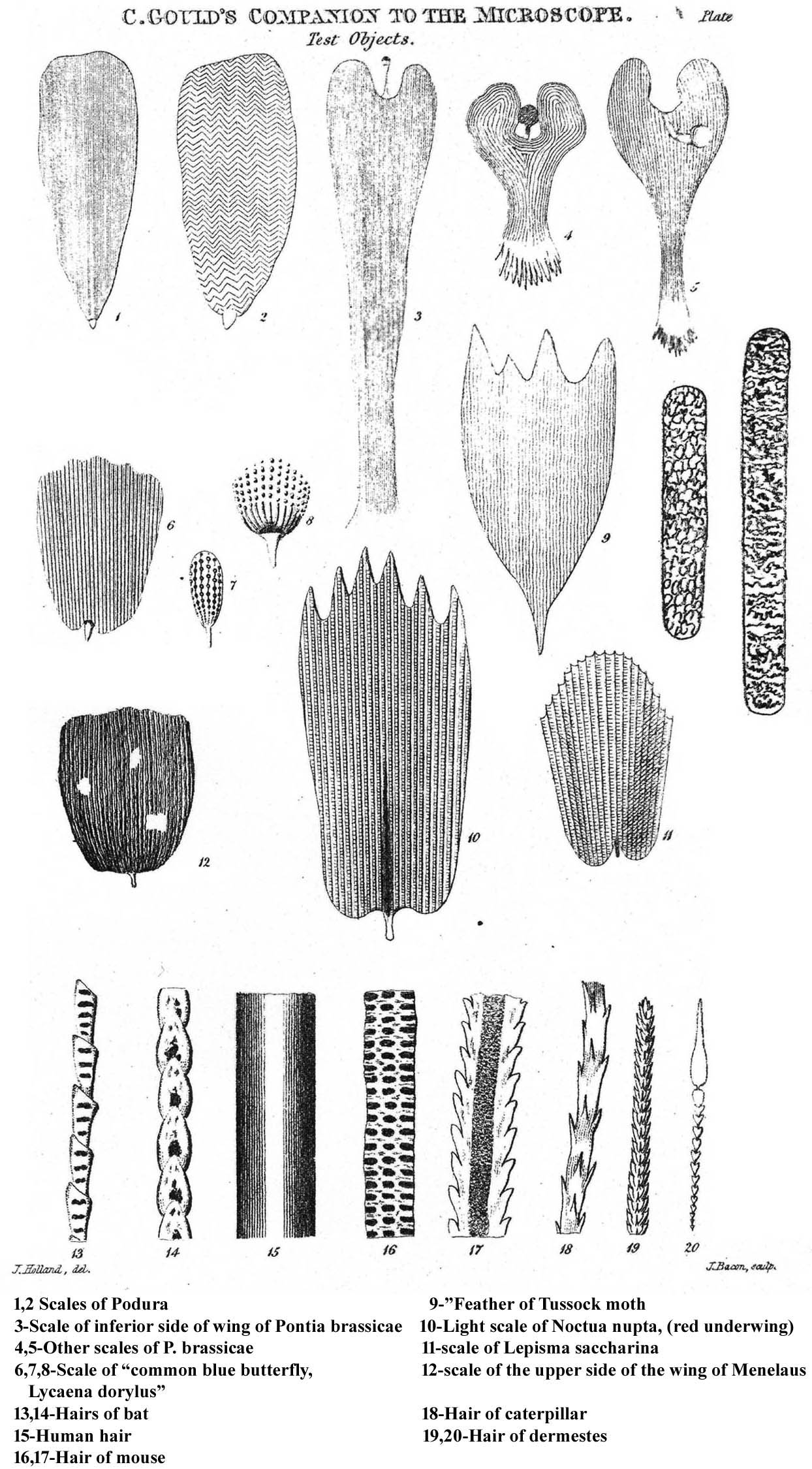
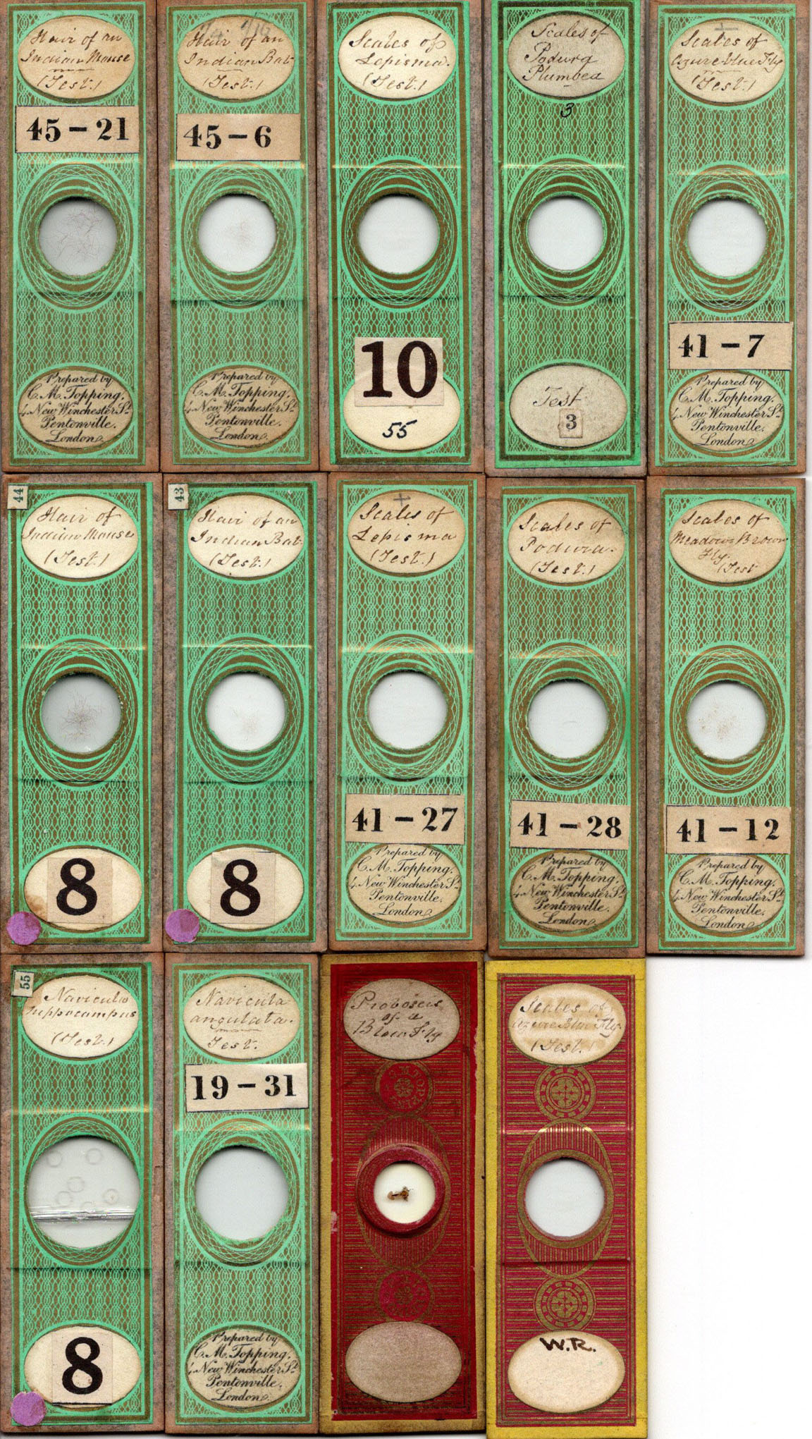
Mr Topping, the excellent preparer of microscopic objects generally furnishes three kinds of test-objects which he carefully covers with the thinnest glass, in order that object glasses of the highest powers may be used to examine them.In this list, there were 9 slides of scales, 6 of hairs, but only only 3 diatoms listed; he stated that the diatoms were much more difficult to
definethan the other objects. He went on to quote Dr Goring
that the study of the manner in which these objects are exhibited is of great importance...for this reason the Infusoria are considered the most valuable and in illuminating them, oblique diverging rays appear to be essential...the degree of obliquity varying with the different specimens.... Topping continued to sell test slides for many years, dying in 1874. Many of his test slides from my collection, are shown to the left.
| OBJECT | IMAGE | OBJECTIVE | EXPECTED FEATURES | ANGULAR APERT | N.A. |
|---|---|---|---|---|---|
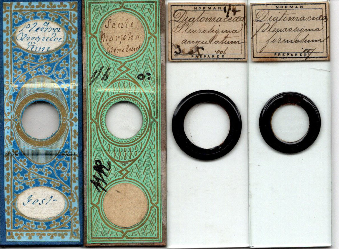
| Scale of Morpho menalus (menalaus) | 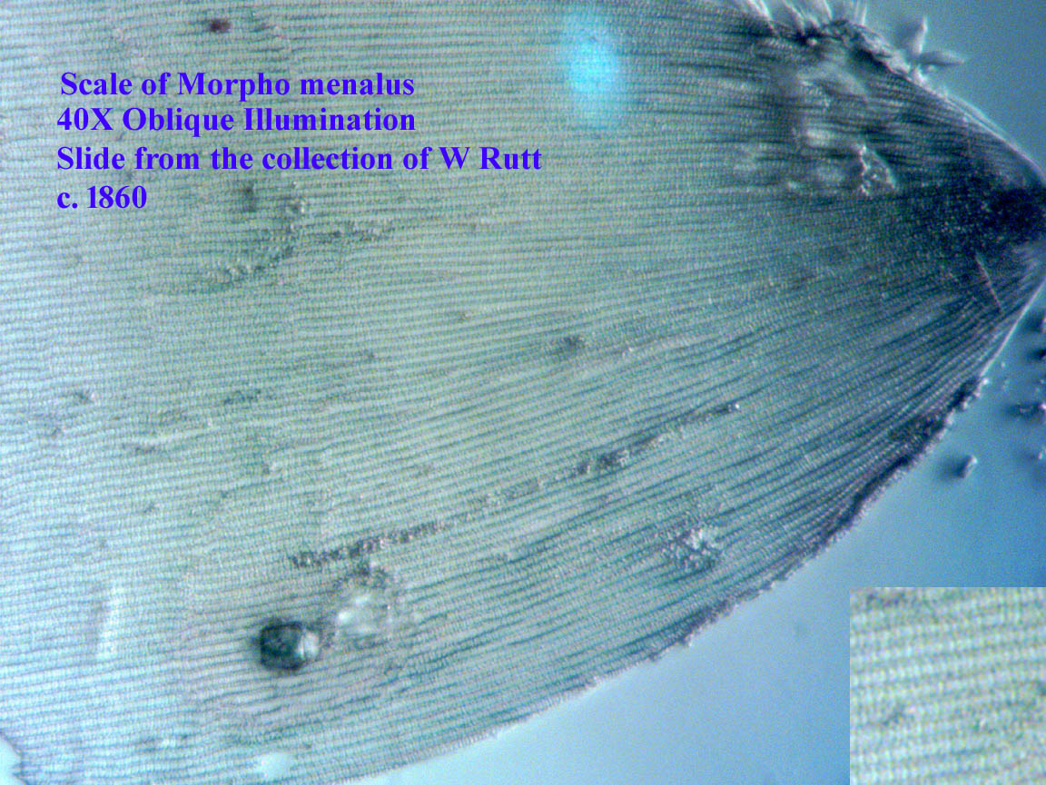 |
Lines will be clear at low powers; Cross markings clear with oblique illumination 40X objective n.a. 0.70. The transverse lines are also visible with oblique top lighting using a lower n.a.; they are clear with oblique top lighting with a 20X 0.46 n.a. objective. The direction of the light is important-it must come from the direction parallel to the long striations | cross striations visible with oblique lighting with n.a. of 0.30 and above | 0.15 to 0.3 | |
Scale of Azure Blue butterfly |
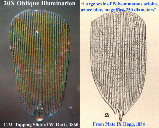 |
Lines will be clear at low powers; Cross markings clear with oblique illumination 20X objective n.a. 0.46. The transverse lines are also visible with oblique top lighting they are clear with oblique top lighting with a 20X 0.46 n.a. objective. The direction of the light is important-it must come from the direction parallel to the long striations | cross markings with n.a. of 0.46 and above | 0.15 to 0.46 | |
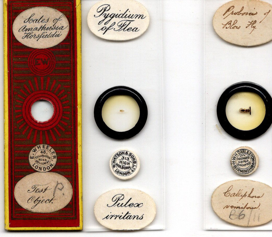
| Probosis of Blowfly | 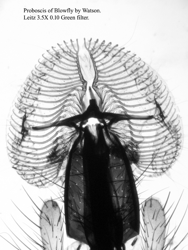 |
Low Powers up to 10X | Thin hairs resolved, edge of coarse hairs distinct wtih a three dimensional effect; periodic structures of spiracles clearly seen, hairs very black, taper to point at ends; must use right side up(hairs should appear first when objective lowered). | 0.15 to 0.3 | |
| Scales of Amathusia horsfieldii | 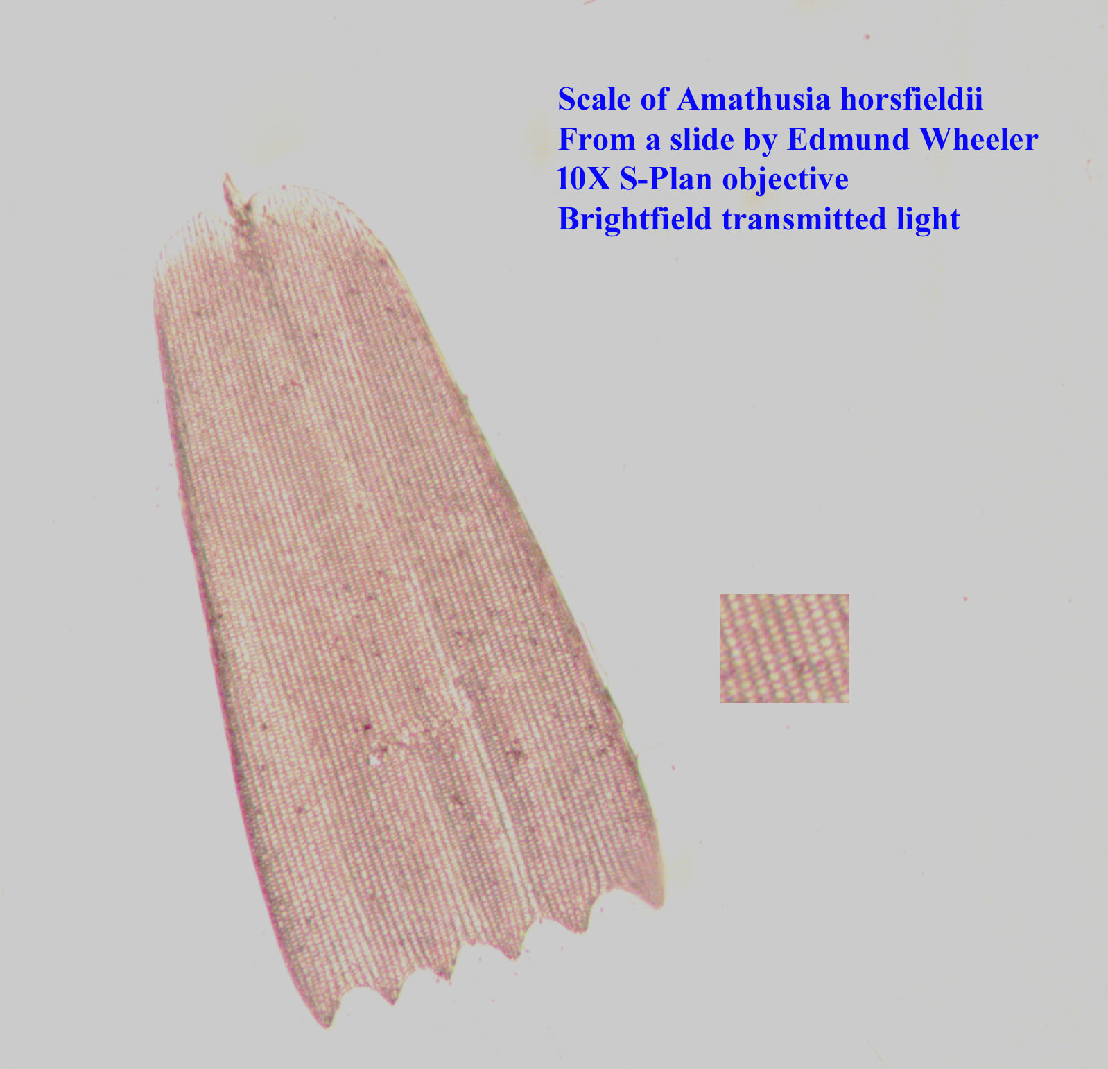 |
10X(?) | Cross striations or beading |
? 0.3 | |
| Scale of Pontia Brassicae(Large White or Cabbage White Butterfly) ( | 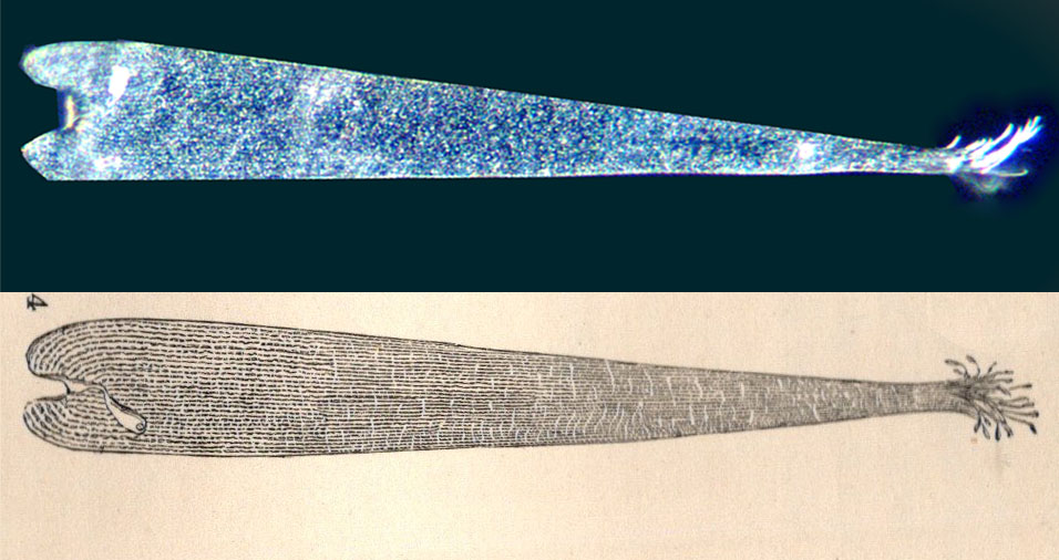 |
Low Powers up to 10X; 20X, n.a. 0.46 reveals the segments well, 40X n.a. 0.70 is needed to see the dots. |
0.15 to 0.70 | ||
| Hair of Indian Bat |  |
Low Powers of a good microscope objective will show the details of overlapping scales and the absence of visible medulla.
The fine hair and the overlapping scales are distinct at 10X. | |||
| Hair of Indian Mouse |  |
Low Powers of a good microscope objective will show the details of the cells in the medulla, the cortex, and the cuticle. It was one of the earliest test objects in the 1820s. The structure varies a bit depending on the thickness of the specimen, the most detail visible in the thickest specimens.
The periodic cellular structure | |||
| A well-stained Histological or Botanical section | 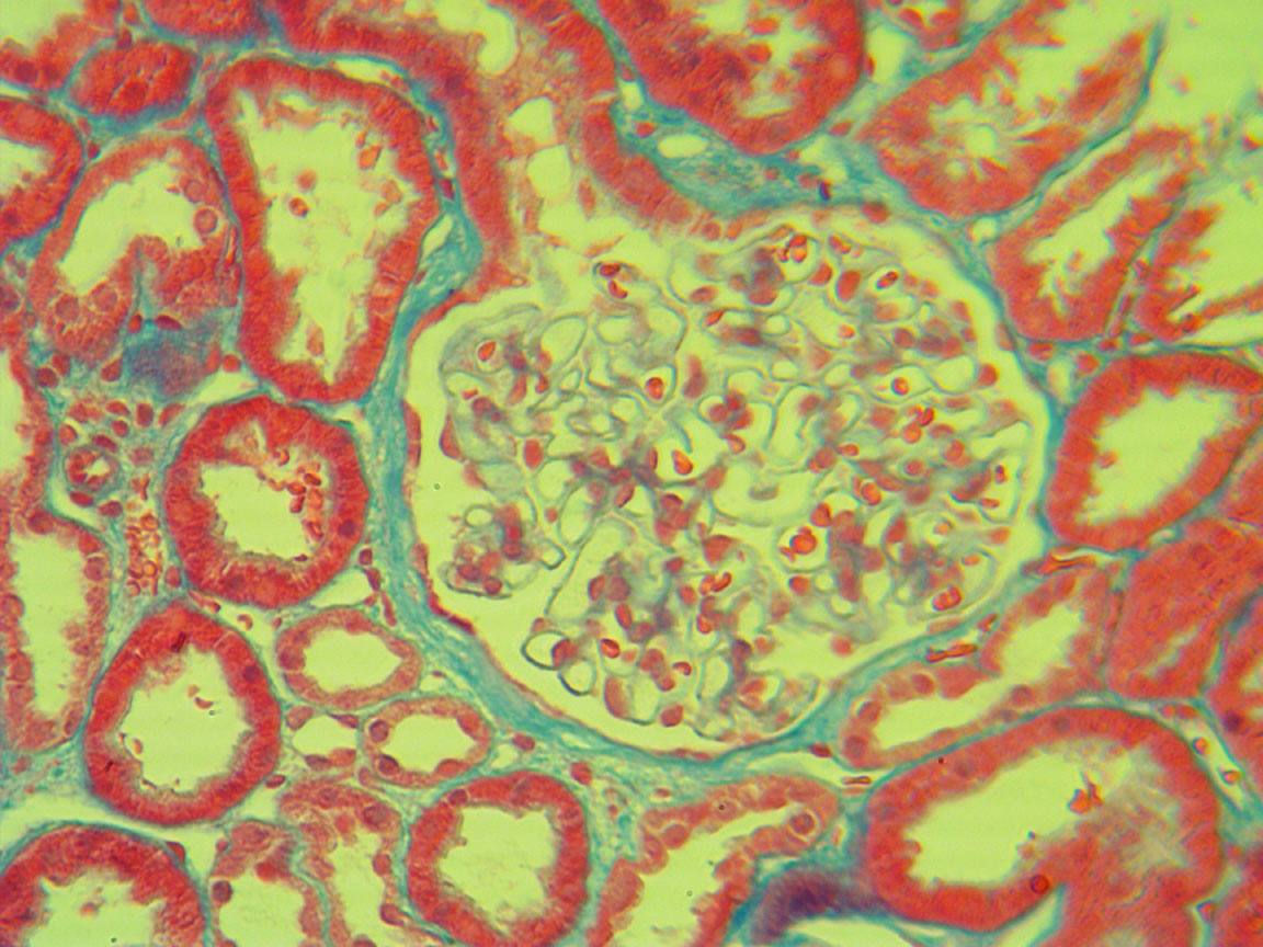 |
Low Powers 3X to 20X | Crisp Image, Structure Well-resolved. The stained image should be brilliant, even with a full cone of light from the substage condenser, especially with a 5X to 10X apochromat. NOTE: best with an evenly cut thin section. | 0.15 to 0.3 | |
| Pygidium of Flea | 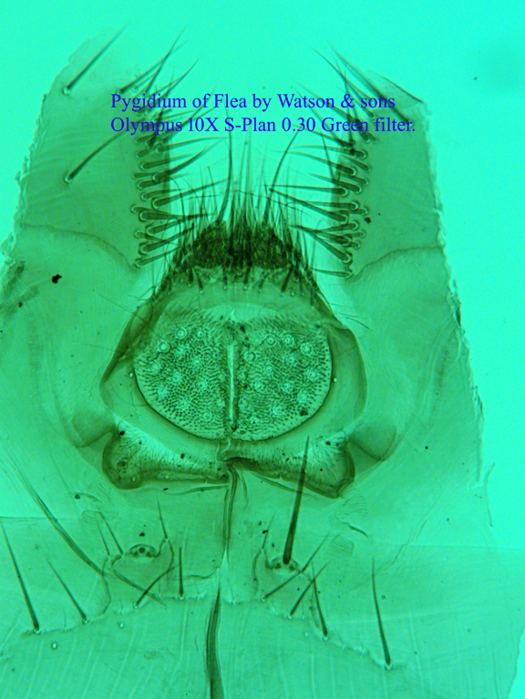 |
10X to 25X | General Outline and Hairs Distinct; define hair implant sites distinctly; substructures clear with na of 0.5 | 22o to 35(50)o | 0.16 to 0.50 |
| Scales of Lepisma saccharina(Silverfish) | 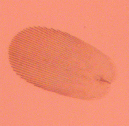 |
10X | lines | Lines are double with clearing in between at 20X with n.a. of 0.46; for more see:David Walker's review of this object | |
| Hemidiscus cuneiformis | 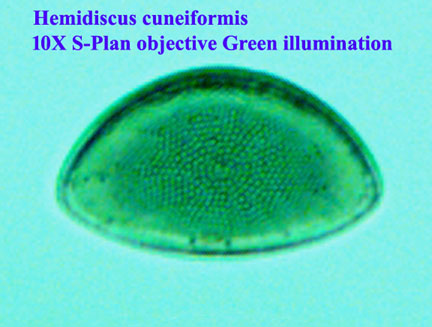 |
10X and up | Resolve areolae with n.a. of 0.30 or more | 0.3 best with green or green & oblique light | |
| Epithemia turgida | 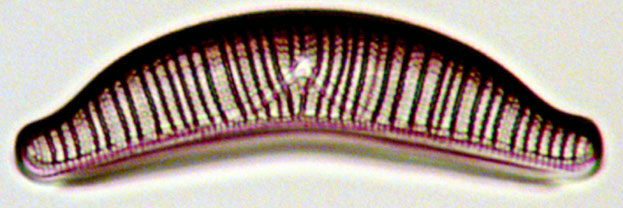 |
10X up; areolae can be discerned at 10X, n.a. 0.30; double rows of areolae clear at 40X with 0.70 n.a. | Good for testing for achromatism single rows of areolae at n.a. 0.30 and double zig-zag areolae become clear at n.a. 0.70. |
0.30 for areolae 0.70 for clear double rows of areolae |
|
| Navicula smithii | 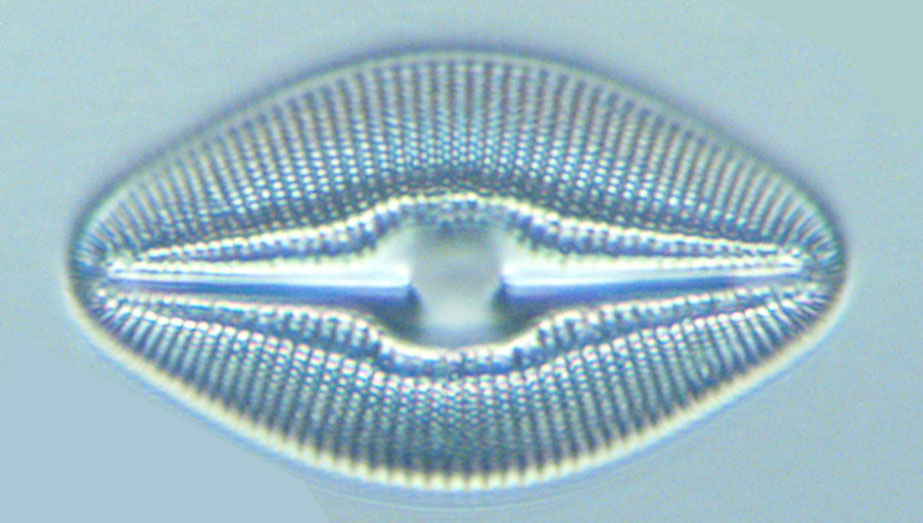 |
20X up | Areolae of this diatom require a n.a. of 0.46 (my 20X S-Plan objective) and oblique illumination perpendicular to the long axis of the valve face; I resolved them into double rows with a 40X S-plan objective n.a. 0.70, again with oblique illumination. | ||
| Frustulia rhomboides (formerly Navicula) | 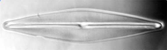 |
According to Hogg, in his 1869 edition, the striae of this diatom, favored by Amici, were considered a very good test of angular aperture. I would agree wholeheartedly, as I could bearly resolve longitudinal striae with my 40X 0.70 objective using oblique illumination. NIC at 40X could resolve some of the areolae. My 100X S-Plan Apo with a n.a. of 1.30 did resolve both striae and areolae very well using oiled darkground illumination. |
over 0.70 | ||
| Cymatopleura soles | 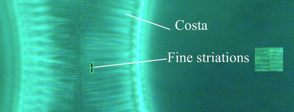 |
40X up | resolution of the fine streae requires very high N.A. | over 1 for the finest striae | |
| Scales of Podura(Springtail) | 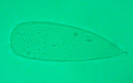 |
10X 0.30 shows dashed appearance; 40X shows the shape of the spines clearly, oil immersion is required to see the hollowed appearance of the spines, best with DIC. | Fine structure of each showing exclamation mark shape and some appearing partly hollowed out is the finding requiring the hihgest resolution, best with DIC and high n.a. e.g. 1.30 | ||
| Hairs of Larva of Dermestes lardarius | 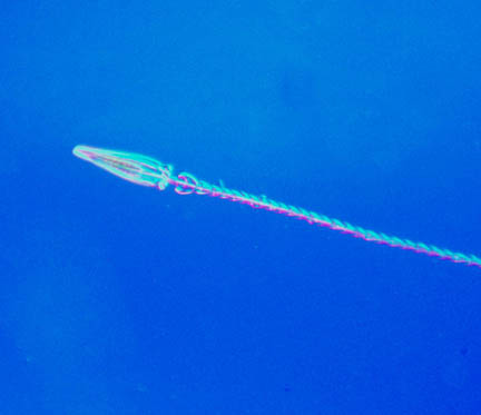 |
Periodic Structures-low power; fine details of the structures-40X | |||
| Mycobacterium Tuberculosis (acid-fast stain) | 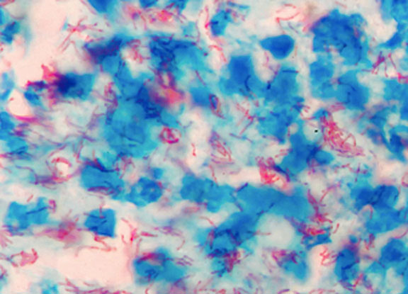 |
Medium to High Dry Objectives | When stained properly, the bacteria should be distinctly red and sharp. Resolution to Beaded appearance; NOTE:This requires a carefully prepared Ziehl-Nielsen Stained specimen, hard to obtain in modern times as most tb specimens are now prepared for florescence; commerical preparations are, as of the time of this writing, of poor quality in the author's experience. | 20X apochromat or high N.A. 30X and above achromats | 0.65 with 15X eyepiece 0.65 to 0.95 n.a. objectives |
| Human Blood Smear-Wright stain or Wright-Giemsa stained(preferred)(NOTE: high quality preparation needed) | 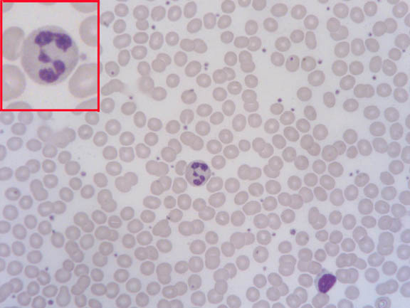 |
20X-60X | Clarity, detail, brilliance and differentiation of staining of the WBCs | 0.65 to 0.95 | |
| Onion Root Tip L.S. stained with iron-hematoxylin | 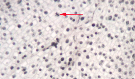 |
Fine test for 20X apo, Higher NA achromats | Mitotic figures black, not gray, spindle fibers discernible. Crisper image with Apo or Flourite | 0.65 to 0.95 | |
| Pleurosigma angulatum | 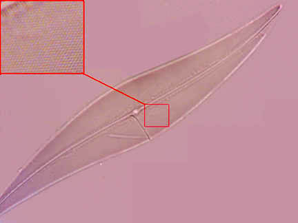 |
White and black dots distinct with 15X eyepiece; with oil immersion objectives, 20X eyepiece and 60X to 120X objectives the black dots are crisp and white ones like brilliant balls without haze-this is also a good test for achromatism-flourites will show slight red and green tints, apochromats almost color-free. | 0.52 with Green light, otherwise 0.85 to 0.95 | ||
| Synedra capitata | 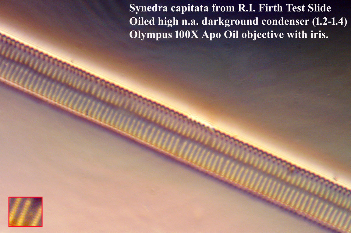 |
90X oil up | Striae with 100X but use of Green oblique illumination or double oiled darkground illumination is usually required for puncta. | ||
| Amphipleura pellucida | 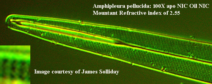 |
High Power Oil Immersion | This is one of the most challenging objects for a light microsope and a specific setup has been recommended. Distinct transverse striae can be seen at 100X with oblique light double oil immersions. Double oil immersion DIC alone has not been successful in my experience especially with mountants with refractive index less than 2. Resolution of the areolae requires oblique monochromatic green or blue light or DIC but only if mounted in Realgar will this work well. I was unable to resolve the areolae except in a Realgar mount with any technique. The direction of the oblique light is very important in the resolution of this diatom. Barnard and Welch stated in the J.R.M.S., volume 51, pages 121-122 in 1932 that clearresolution of the areolae would require ultraviolet oblique or darkground illumination. |
1.25 to 1.4 |