DIATOMS
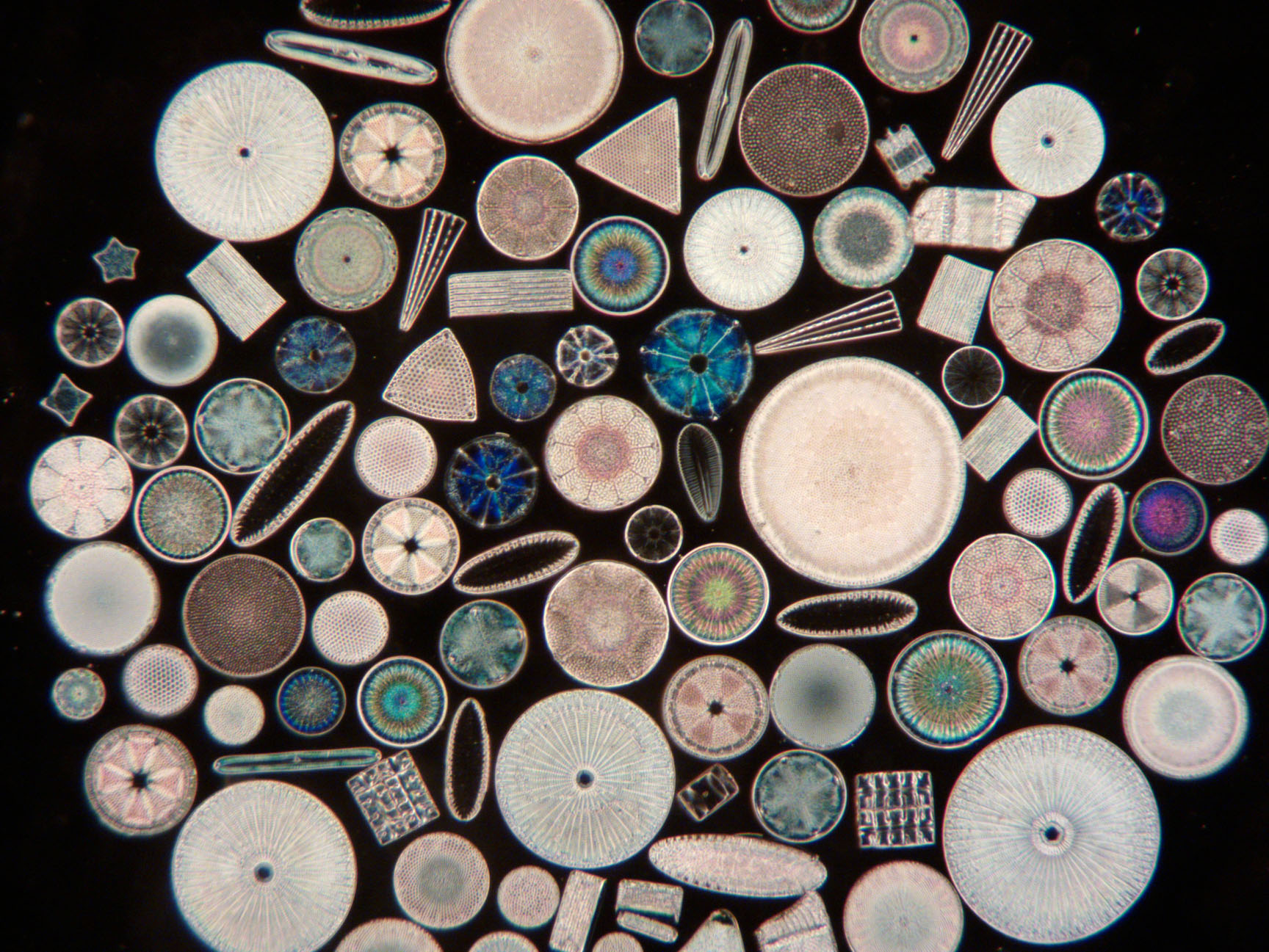
Diatoms are a diverse group of unicellular algae found in oceans, freshwater, and even moist soils. There are at least 30,000 known species and the number of species may number in the hundreds of thousands. They evolve rapidly, have wide genetic diversity and have evolved to adapt to dramatically different environments. They are a critical part of aquatic ecosystems and play a vital role in global cycles of carbon and oxygen because of their process of photosynthesis. Diatoms have a rigid cell wall, called a frustule, with intricate patterns of hydrated silicon dioxide (silica), earning them the nickname "glass houses". This frustule remains after the diatom dies, forming deposits known as diatomaceous earth. Diatoms have a wide range of shapes and sizes, from 2 to 500 microns (micrometers). Individual diatoms vary tremendously in shape and these shapes include cylinders, pointed stars, crosses, cubes, cigar shapes, S-shapes, triangles etc. They can be solitary or form colonies with varied structures such as ribbons, fans, zigzags, or stars. In summary, diatoms are amazing microscopic organisms that play a fundamental role in global ecosystems, acting as oxygen producers, primary producers, and indicators of environmental health. Many have strikingly beautiful structures.
Diatoms are usually symmetrical in at least one dimension. Their siliceous skeletons are referred to as frustules. The diatoms use silicic acid to create their frustules. This soluble form of silica is taken up by silicon transporters into the silica deposition vesicle where it is precipitated. Several molecules have been implicated in the precipitation and structure on a nanoscale. These include silaffins, silacidins, cingulins and long-chain polyamines. Evidence suggests that structuring of assemblages and the final frustule shape is influenced by actin microfilaments and microtubules of the cytoskeleton.
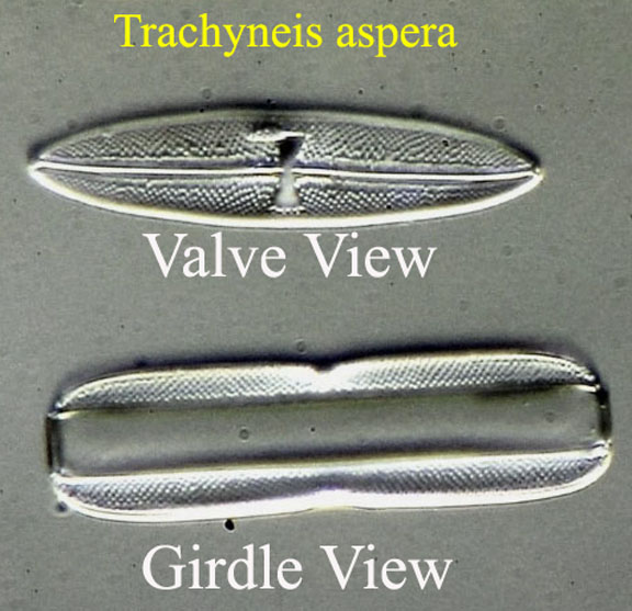
In the grossest terms, a diatom is a box-shaped organism, the top of which is wider than the side. The top is called the valve and the side the girdle.
Diatoms can be seen under the microscope from the valve view or girdle view. In most cases, diatoms are mounted in valve view because they naturally often settle this way when placed on a slide. In some cases, they are mounted in girdle view which is in most cases more difficult to accomplish on a slide. Much less commonly, both views are provided. Examining the diatom in only one view can be misleading in terms of the overall three dimensional shape as is shown by the view of both from the same slide shown here.
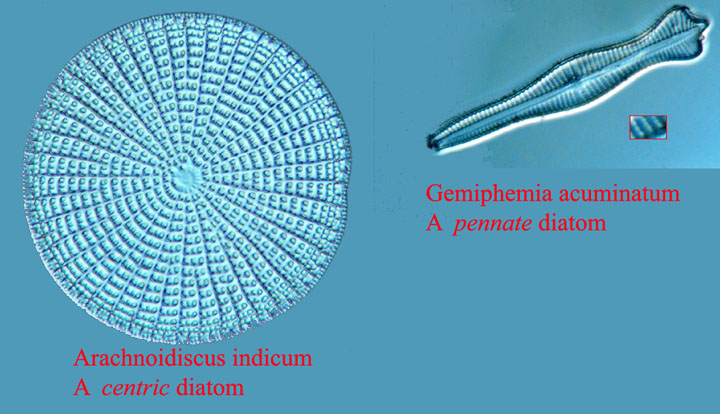
Diatoms are mainly categorized into two groups based on the symmetry of their frustules:
Centric Diatoms: These have radial symmetry, appearing circular, triangular, or polygonal in shape.
Pennate Diatoms: These display bilateral symmetry, typically elongated and possessing a raphe, a slit-like structure used for gliding movement.
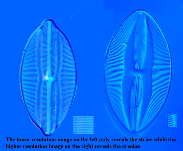
Diatoms can have a very complex structure. Various structures have been assigned different names, which are at times confusing. Among the most interesting and important are the perforations which may be referred to as puncta. The smaller and more numerous perforations are known as areolae and are usually best seen in valve view. These perforations vary in size and also in periodicity. In most cases, people looking at diatoms under the microscope are interested in periodic structures. Among the most commonly noted periodic structures are the striae and areolae. Striae are lines that under higher magnification and resolving power, are found to be composed of dots
which are the areolae. The term puncta simply refers to any perforation and includes the areolae. The spacing between each areola varies with the species. The visibility of areola puncta in different species of diatoms have therefore been used as tests of different degrees of resolution. A page showing an 8 form test slide by the late Klaus Kemp is on this site, as are others. In some cases, puncta which are not areolae have a periodic structure, which can be both confusing and misleading. Furthermore, exact spacing of the areolae may vary even in the same species, though not usually markedly.
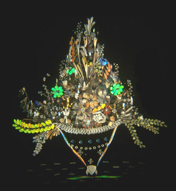
In addition to use as test objects, diatoms have sometimes been used to create complex images under the microscope, either composed of only diatoms, or combined with the scales of butterfly wings. These images are referred to as arrangements
or arranged
diatoms. These arrangements were produced in the 19th century as an entertainment for microscopists, and in the 20th century by Klaus Kemp of England (d. 2022), and others. Today there are still a few mounters who practice this art. Antique as well as modern examples were quite tedious to produce and are usually quite expensive today, regardless of age.
Slides of diatoms may be strews where a variety are randomly thrown
on to a slide, often with some diatoms overlapping others. These slides can provide a great variety of forms, but often include parts of diatoms, overlapping diatoms, and sometimes contaminants. In other cases slides have been made of a single species, or a group of related species carefully placed on the slide. In some cases multiple types are carefully placed one at a time on a type slide which may contain tens, hundreds or even thousands of different species. In some cases, a list accompanies the slide, as is the case with the 37-diatom type slide by N.B.S. in this collection, and a 100-form type slide by Klaus Kemp, also in this collection. In other cases, as the 530 form Kemp slide in this collection, no list was provided, leaving the user to identify them. Less common are slides in which the diatoms are mounted in a microphotographic grid with each identified with its name visible under the diatom under the microscope. An example of one of these is the Moller 335 type slide in this collection. In general the more forms on the type slide, the more valuable the slide. In the past, slides containing thousands of diatoms were made by famous mounters like Moller. These were special order slides, provided only on request, (and at great expense) to selected individuals such as the famous diatomist Henri van Heurck. Moller and Thum routinely made type slides of hundreds of diatoms. They referred to such slides as Typen-platte
.
Obtaining Diatom Slides:
In addition to antique slides sold on Ebay, as of 2025, new diatom slides are still being sold by S. Barone at diatomshop.com. High quality slides mounted by the late Klaus D. Kemp were made in the late 20th through the early 21st century and are of good quality; their only negative is that except on special request, diatoms were mounted on the slide with the coverslip mounted on top; he only mounted on the coverslip by special request. S. Barone currently makes very high quality slides, with the diatoms mounted on the underside of the coverslip rather than the slide, using moderately high refractive index mountant, and nicely sealed for preservation. This method, while tedious, and therefore expensive, provides the best resolution of details with the light microscope as used today. Unfortunately slides of some important diatoms, like Amphipleura pellucida,as of this writing(2025), are not available from diatomshop.com.
Resolving the areolae have been an obsession with microscopists for centuries. In many cases, this requires an objective with high numerical aperture (n.a.), and oil immersion of both the objective and the high n.a. condenser. For many difficult
ones, where the distance between areolae is small, special techniques are required such as oblique lighting, D.I.C., phase contrast or dark ground illumination. In my experience, oblique lighting using a dichroic filter at the optimal wavelength for the objective is most often successful, though one needs to be able to rotate the direction of the lighting to suit the individual diatom. Taking images with a camera and stacking the images may improve the clarity of the areolae. Occasionally dark ground or one of the other techniques works better. Lastly, for diatoms in which the areolae spacing are close to the limit of the light microscope, high refractive index (r.i.) mountant may be needed to resolve the areolae. For the last 75 years or so, the two mountants commonly used for diatoms have been Styrax and Hyrax. Styrax has a r.i. of about 1.6 whereas Hyrax is about 1.7 and Nephrax is >1.75
. The last is likely the highest r.i. mountant still available. In the past, slides were made with an even higher refractive index mountant, Realgar (arsenic sulfide, r.i. 2-2.5), but this is no longer used as the process is hazardous. Occasionally antique Realgar slides come to market, and these may provide an advantage over the newer slides IF one is lucky enough to have one with the specimens mounted close to, or on the bottom of, the coverslip.
It should be noted that the relatively inexpensive C-mount microscope cameras of today do not have the high resolution provided by the film and camera arrangements of the past as used by such people as Spitta, so duplicating some of the best images by Spitta may not be possible with these. Higher resolution microscope cameras are available. I use a C-mount camera that cost about $1000 (2025) which has larger pixels (2.9 microns), and built-in software with HDMI output directly to my monitor which is both easier to use and gives somewhat better images than PC-based software used with cheaper C-mount cameras which often have smaller pixels which reduces resolution of the image. Professional microscope cameras often cost much more, but will give a better image with less "noise" and less pixelation, but few of us can afford such expensive devices.